Division of Orthopaedics, Department of Trauma and Orthopaedic Surgery, University Medical Center Hamburg-Eppendorf, Martinistraße 52, 20246, Hamburg, Germany.
Calcif Tissue Int. 2024 Aug;115(2):142-149. doi: 10.1007/s00223-024-01237-w. Epub 2024 Jun 4.
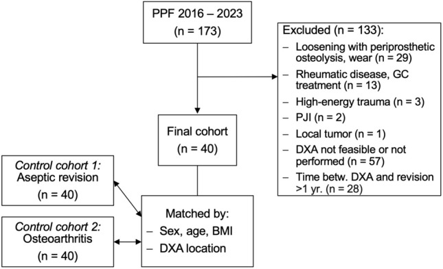
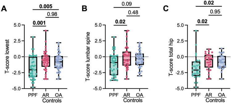
Periprosthetic femoral hip fractures are subject to an increasing incidence and are often considered to be related to osteoporosis. However, there are no available studies that have determined the frequency of osteoporosis in affected patients using gold standard dual-energy X-ray absorptiometry (DXA). In this retrospective comparative study, we analyzed the DXA results of 40 patients with periprosthetic femoral hip fractures who were treated surgically in our department. DXA measurements were performed at the total hip and the lumbar spine to determine bone mineral density T-scores. Data were compared to two age-, sex-, and BMI-matched control groups in which patients underwent DXA prior to aseptic revision surgery for other causes or primary THA (consisting of 40 patients each). The mean T-score in the periprosthetic fracture cohort was significantly lower (- 1.78 ± 1.78) than that of the aseptic revision (- 0.65 ± 1.58, mean difference - 1.13 [95% CI - 1.88 to - 0.37]; p = 0.001) and the primary THA cohort (- 0.77 ± 1.34, mean difference - 1.01 [95% CI - 1.77 to - 0.26]; p = 0.005). Accordingly, osteoporosis was detected more frequently (45%) in the fracture cohort compared to patients undergoing aseptic revision (12.5%) and primary THA (10%). In conclusion, almost half of the patients with periprosthetic femoral hip fractures have osteoporosis according to DXA measurements. A regular assessment of bone health in THA enables identification of patients with osteoporosis who likely benefit from initiation of osteoporosis medication and cemented stem fixation.
人工髋关节假体周围股骨骨折的发病率不断上升,通常认为与骨质疏松症有关。然而,目前尚无研究使用金标准双能 X 射线吸收法(DXA)确定受影响患者骨质疏松症的频率。在这项回顾性对比研究中,我们分析了在我院接受手术治疗的 40 例人工髋关节假体周围股骨骨折患者的 DXA 结果。DXA 测量在全髋关节和腰椎进行,以确定骨矿物质密度 T 评分。将数据与因其他原因接受无菌翻修手术或初次全髋关节置换术(各包含 40 例患者)的两个年龄、性别和 BMI 匹配的对照组进行比较。假体周围骨折组的平均 T 评分明显低于无菌翻修组(-1.78±1.78)(-0.65±1.58,平均差异-1.13[95%CI-1.88 至-0.37];p=0.001)和初次全髋关节置换组(-0.77±1.34,平均差异-1.01[95%CI-1.77 至-0.26];p=0.005)。因此,与无菌翻修组(12.5%)和初次全髋关节置换组(10%)相比,骨折组中骨质疏松症的检出率更高(45%)。总之,根据 DXA 测量,近一半的人工髋关节假体周围股骨骨折患者患有骨质疏松症。对全髋关节置换患者的骨骼健康进行常规评估,可以发现患有骨质疏松症的患者,他们可能受益于开始骨质疏松症药物治疗和骨水泥固定。