Department of Dermatology, Seoul National University College of Medicine, Seoul, Republic of Korea.
Institute of Human-Environment Interface Biology, Medical Research Center, Seoul National University College of Medicine, Seoul, Republic of Korea.
Front Immunol. 2024 May 22;15:1365430. doi: 10.3389/fimmu.2024.1365430. eCollection 2024.
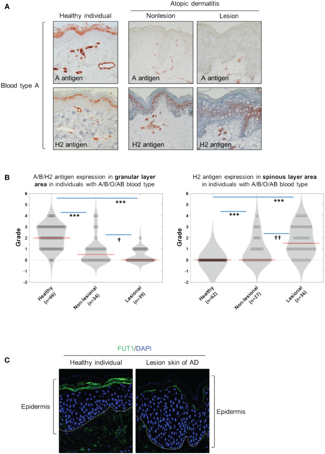
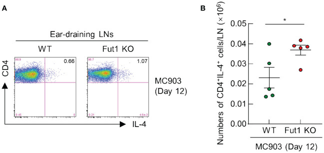
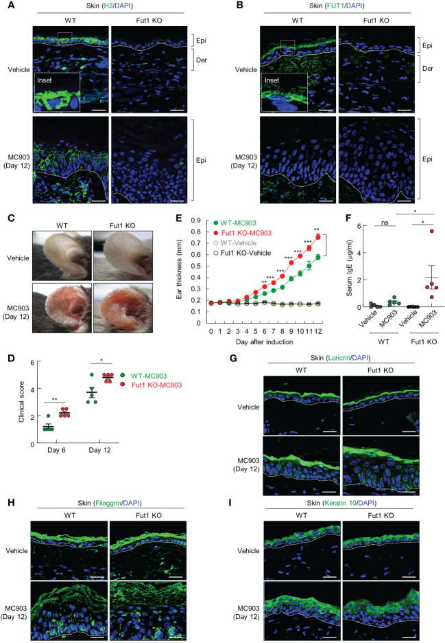
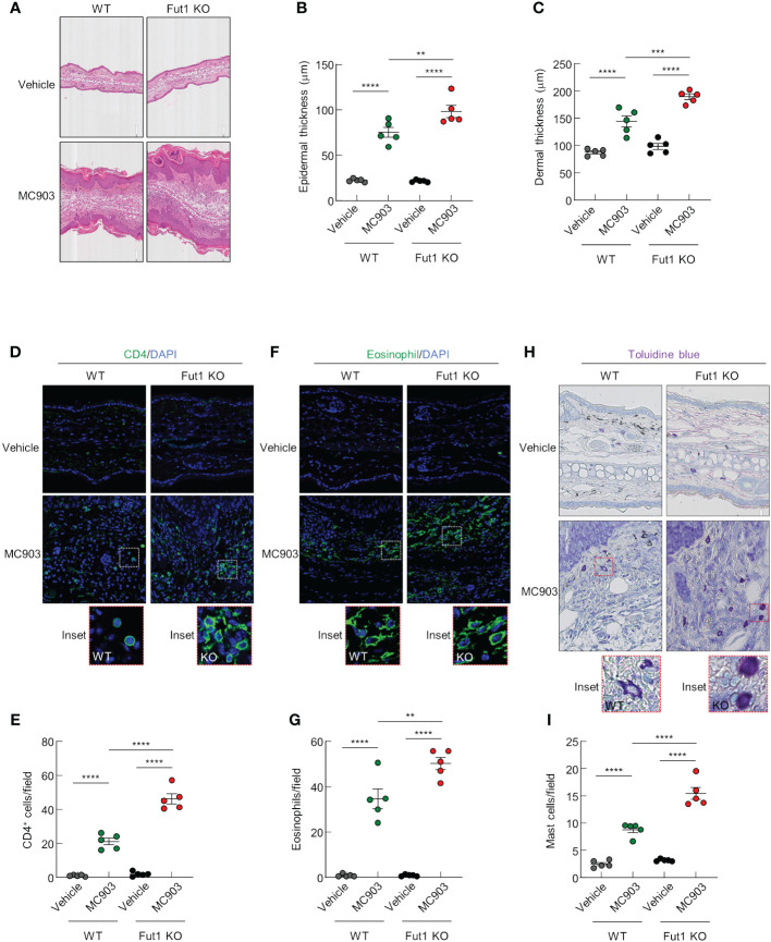
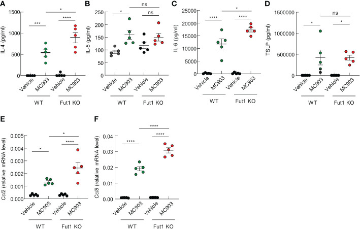
The presence of the blood group H2 antigen on the membrane of red blood cells determines blood type O in individuals and this H2 antigen serves as a precursor to the A and B antigens expressed in blood types A and B, respectively. However, the specific involvement of ABH antigens in skin diseases is unknown. Therefore, we aim to investigate the expression of ABH antigens in skin tissue of patients with atopic dermatitis (AD) and MC903-induced AD-like mice. We demonstrated that the expression of ABH antigen is primarily located in the granular and horny layers of the skin in healthy control individuals. However, in patients with AD, the expression of the ABH antigen was absent or diminished in these layers, while the H2 antigen expression increased in the spinous layers of the affected skin lesions. Then, we investigated the biological function of blood group H antigen mediated by fucosyltransferase 1 (Fut1) in the skin, utilizing an AD mouse model induced by MC903 in wild-type (WT) and -knockout mice. After the application of MC903, Fut1-deficient mice, with no H2 antigen expression on their skin, exhibited more severe clinical signs, increased ear swelling, and elevated serum IgE levels compared with those of WT mice. Additionally, the MC903-induced thickening of both the epidermis and dermis was more pronounced in Fut1-deficient mice than that in WT mice. Furthermore, Fut1-deficient mice showed a significantly higher production of interleukin-4 (IL-4) and IL-6 in skin lesions compared with that of their WT counterparts. The expression of chemokines, particularly and , was notably higher in Fut1-deficient mice compared with those of WT mice. The infiltration of CD4 T cells, eosinophils, and mast cells into the lesional skin was significantly elevated in Fut1-deficient mice compared with that in WT mice. These findings demonstrate the protective role of H2 antigen expression against AD-like inflammation and highlight its potential therapeutic impact on AD through the regulation of blood group antigens.
红细胞膜上 H2 抗原的存在决定了个体的血型为 O 型,而这种 H2 抗原分别作为血型 A 和 B 中 A 抗原和 B 抗原的前体。然而,ABH 抗原在皮肤病中的具体作用尚不清楚。因此,我们旨在研究特应性皮炎(AD)患者和 MC903 诱导的 AD 样小鼠皮肤组织中 ABH 抗原的表达。我们发现,ABH 抗原在健康对照个体的皮肤颗粒层和角质层中主要表达。然而,在 AD 患者中,这些层的 ABH 抗原表达缺失或减少,而受影响皮肤病变的棘层中 H2 抗原表达增加。然后,我们利用 MC903 诱导的 AD 小鼠模型,研究了皮肤中糖基转移酶 1(Fut1)介导的血型 H 抗原的生物学功能。在 Fut1 缺陷型(无 H2 抗原表达)和野生型(WT)小鼠中,应用 MC903 后,前者的临床症状更严重,耳部肿胀增加,血清 IgE 水平升高。此外,与 WT 小鼠相比,Fut1 缺陷型小鼠的表皮和真皮均明显增厚。此外,Fut1 缺陷型小鼠皮肤病变中白细胞介素-4(IL-4)和白细胞介素-6(IL-6)的产生显著高于 WT 小鼠。趋化因子的表达,特别是 和 ,在 Fut1 缺陷型小鼠中明显高于 WT 小鼠。Fut1 缺陷型小鼠病变皮肤中 CD4 T 细胞、嗜酸性粒细胞和肥大细胞的浸润显著高于 WT 小鼠。这些发现表明 H2 抗原表达对 AD 样炎症具有保护作用,并强调了其通过调节血型抗原对 AD 的潜在治疗作用。