• 文献检索
  • 文档翻译
  • 深度研究
  • 学术资讯
  • Suppr Zotero 插件Zotero 插件
  • 邀请有礼
  • 套餐&价格
  • 历史记录
应用&插件
Suppr Zotero 插件Zotero 插件浏览器插件Mac 客户端Windows 客户端微信小程序
定价
高级版会员购买积分包购买API积分包
服务
文献检索文档翻译深度研究API 文档MCP 服务
关于我们
关于 Suppr公司介绍联系我们用户协议隐私条款
关注我们

Suppr 超能文献

核心技术专利:CN118964589B侵权必究
粤ICP备2023148730 号-1Suppr @ 2026

文献检索

告别复杂PubMed语法,用中文像聊天一样搜索,搜遍4000万医学文献。AI智能推荐,让科研检索更轻松。

立即免费搜索

文件翻译

保留排版,准确专业,支持PDF/Word/PPT等文件格式,支持 12+语言互译。

免费翻译文档

深度研究

AI帮你快速写综述,25分钟生成高质量综述,智能提取关键信息,辅助科研写作。

立即免费体验

犹豫不决:联合多模态成像与腹腔镜检查诊断结核性腹膜炎

On the fence: Combining multimodal imaging and laparoscopy for diagnosing tuberculous peritonitis.

作者信息

Yamada Kenji, Aoki Kazuaki, Tanaka Rina, Matsushima Seito, Sato Akiyuki, Kobayashi Takaaki, Moody Sandra, Nogi Masayuki

机构信息

Department of General Internal Medicine Kameda Medical Center Kamogawa, Chiba Japan.

Division of Infectious Diseases University of Iowa Iowa City Iowa USA.

出版信息

Clin Case Rep. 2024 Jun 5;12(6):e8996. doi: 10.1002/ccr3.8996. eCollection 2024 Jun.

DOI:10.1002/ccr3.8996
PMID:38845802
原文链接:https://pmc.ncbi.nlm.nih.gov/articles/PMC11154765/
Abstract

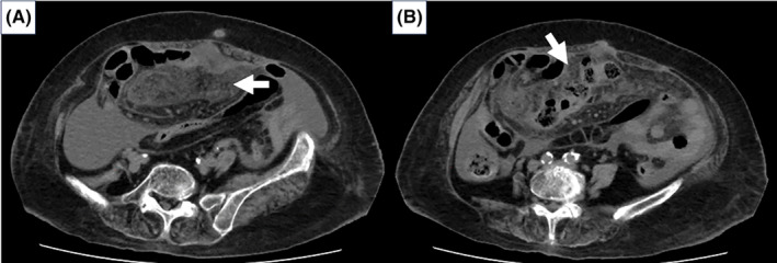
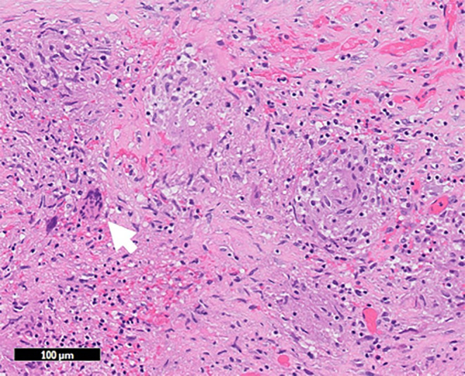
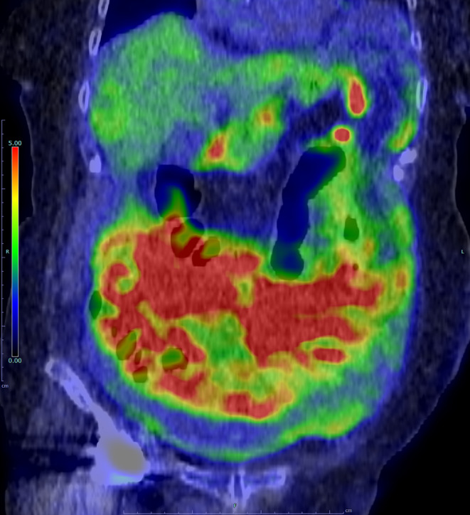
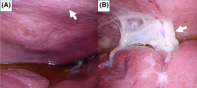
Tuberculous peritonitis (TB peritonitis) is one of the most challenging forms of extrapulmonary TB to diagnose. While tumor markers can be elevated in patients with TB peritonitis, FDG-PET/CT can aid in distinguishing TB peritonitis from malignancies, if an apron-like omentum pattern is seen. Laparoscopy is crucial for accurate and early diagnosis.

摘要

结核性腹膜炎是肺外结核中最难诊断的类型之一。虽然结核性腹膜炎患者的肿瘤标志物可能升高,但如果出现围裙样大网膜模式,氟脱氧葡萄糖正电子发射断层扫描/计算机断层扫描(FDG-PET/CT)有助于鉴别结核性腹膜炎与恶性肿瘤。腹腔镜检查对准确早期诊断至关重要。

https://cdn.ncbi.nlm.nih.gov/pmc/blobs/9ea3/11154765/e417cbdfdf7d/CCR3-12-e8996-g001.jpg
https://cdn.ncbi.nlm.nih.gov/pmc/blobs/9ea3/11154765/1421e153da5e/CCR3-12-e8996-g004.jpg
https://cdn.ncbi.nlm.nih.gov/pmc/blobs/9ea3/11154765/1434fbde17c7/CCR3-12-e8996-g003.jpg
https://cdn.ncbi.nlm.nih.gov/pmc/blobs/9ea3/11154765/425a017c1827/CCR3-12-e8996-g002.jpg
https://cdn.ncbi.nlm.nih.gov/pmc/blobs/9ea3/11154765/e417cbdfdf7d/CCR3-12-e8996-g001.jpg

相似文献

1
On the fence: Combining multimodal imaging and laparoscopy for diagnosing tuberculous peritonitis.犹豫不决:联合多模态成像与腹腔镜检查诊断结核性腹膜炎
Clin Case Rep. 2024 Jun 5;12(6):e8996. doi: 10.1002/ccr3.8996. eCollection 2024 Jun.
2
Characterizing omental PET/CT findings for differentiating tuberculous peritonitis from peritoneal carcinomatosis.描述网膜 PET/CT 的表现,以区分结核性腹膜炎与腹腔肿瘤转移。
Abdom Radiol (NY). 2021 Dec;46(12):5574-5585. doi: 10.1007/s00261-021-03286-3. Epub 2021 Sep 21.
3
[18F-fluorodeoxyglucose-positron emission tomography (FDG-PET) was useful tool for detecting tuberculous peritonitis: report of a case].[18F-氟脱氧葡萄糖正电子发射断层扫描(FDG-PET)是检测结核性腹膜炎的有用工具:病例报告]
Nihon Shokakibyo Gakkai Zasshi. 2008 Oct;105(10):1515-22.
4
A case report of peritoneal tuberculosis diagnosed by laparoscopy in a low prevalence setting.腹腔镜诊断低流行地区腹膜结核 1 例报告。
Acta Chir Belg. 2021 Aug;121(4):269-273. doi: 10.1080/00015458.2019.1693152. Epub 2019 Nov 24.
5
Advances in imaging of tuberculosis: the role of ¹⁸F-FDG PET and PET/CT.结核病影像学进展:¹⁸F-FDG PET 和 PET/CT 的作用。
Curr Opin Pulm Med. 2014 May;20(3):287-93. doi: 10.1097/MCP.0000000000000043.
6
Tuberculous peritonitis--do not miss it.结核性腹膜炎——切勿漏诊。
Dis Colon Rectum. 1996 Apr;39(4):394-9. doi: 10.1007/BF02054053.
7
Value of gamma interferon enzyme-linked immunospot assay in the diagnosis of peritoneal dialysis-associated tuberculous peritonitis.γ干扰素酶联免疫斑点试验在诊断腹膜透析相关性结核性腹膜炎中的价值。
Int Urol Nephrol. 2022 Apr;54(4):843-849. doi: 10.1007/s11255-021-02960-1. Epub 2021 Jul 14.
8
[Tuberculous peritonitis. 'Forgotten' disease].[结核性腹膜炎。“被遗忘的”疾病]
Khirurgiia (Mosk). 2018(12):38-44. doi: 10.17116/hirurgia201812138.
9
Pretreatment CT differential diagnosis of tuberculous peritonitis from peritoneal carcinomatosis of advanced epithelial ovarian cancer.术前 CT 鉴别诊断结核性腹膜炎与晚期上皮性卵巢癌腹膜转移。
Sci Rep. 2023 Jan 19;13(1):1085. doi: 10.1038/s41598-023-27771-5.
10
Imaging in tuberculosis.结核病影像学诊断。
Int J Infect Dis. 2015 Mar;32:87-93. doi: 10.1016/j.ijid.2014.12.007.

本文引用的文献

1
An Updated Systematic Review and Meta-Analysis for the Diagnostic Test Accuracy of Ascitic Fluid Adenosine Deaminase in Tuberculous Peritonitis.结核性腹膜炎中腹水腺苷脱氨酶诊断试验准确性的最新系统评价和荟萃分析
Infect Chemother. 2023 Jun;55(2):264-277. doi: 10.3947/ic.2023.0014.
2
Characterizing omental PET/CT findings for differentiating tuberculous peritonitis from peritoneal carcinomatosis.描述网膜 PET/CT 的表现,以区分结核性腹膜炎与腹腔肿瘤转移。
Abdom Radiol (NY). 2021 Dec;46(12):5574-5585. doi: 10.1007/s00261-021-03286-3. Epub 2021 Sep 21.
3
Systematic review: tuberculous peritonitis--presenting features, diagnostic strategies and treatment.
系统评价:结核性腹膜炎——临床表现、诊断策略及治疗
Aliment Pharmacol Ther. 2005 Oct 15;22(8):685-700. doi: 10.1111/j.1365-2036.2005.02645.x.