Seidemann Suse, Salomon Florian, Hoffmann Karl B, Kurth Thomas, Sbalzarini Ivo F, Haase Robert, Ader Marius
Center for Regenerative Therapies Dresden (CRTD), Technische Universität Dresden, Dresden, Germany.
Center for Molecular and Cellular Bioengineering (CMCB), Technische Universität Dresden, Dresden, Germany.
Front Mol Neurosci. 2024 May 24;17:1398447. doi: 10.3389/fnmol.2024.1398447. eCollection 2024.
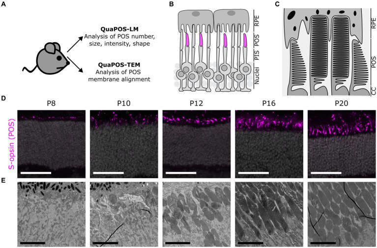
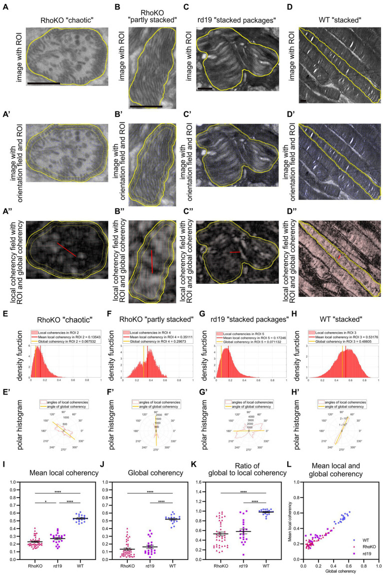
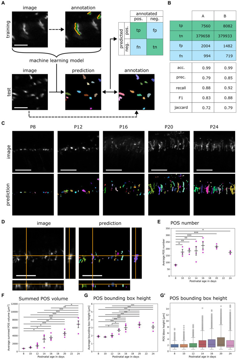
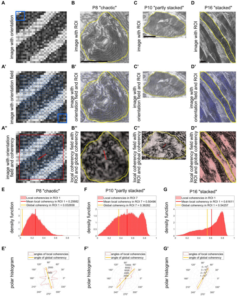
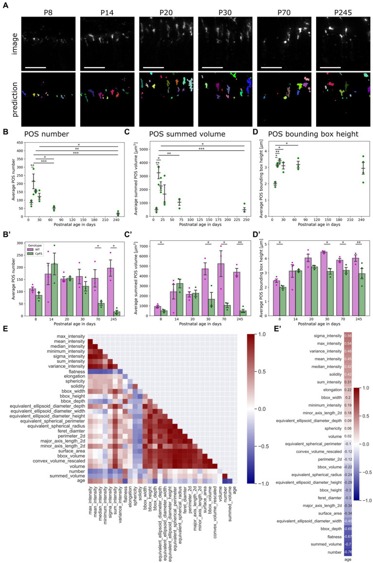
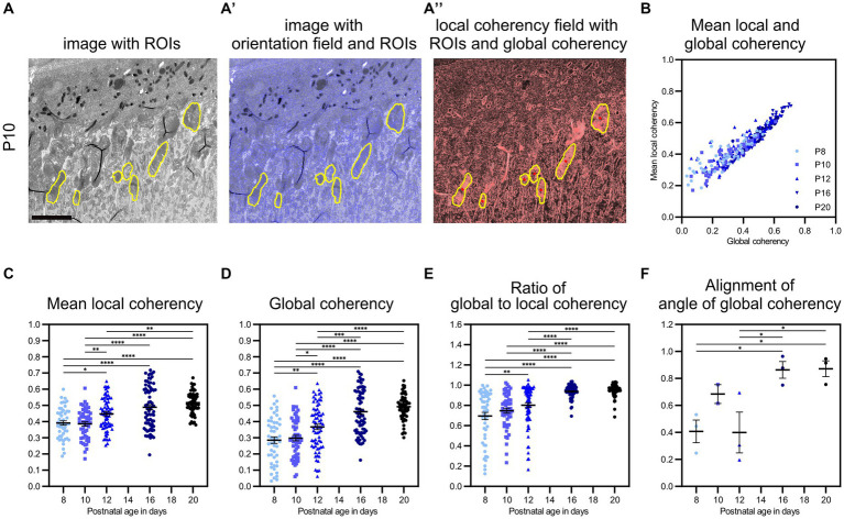
The functionality of photoreceptors, rods, and cones is highly dependent on their outer segments (POS), a cellular compartment containing highly organized membranous structures that generate biochemical signals from incident light. While POS formation and degeneration are qualitatively assessed on microscopy images, reliable methodology for quantitative analyses is still limited. Here, we developed methods to quantify POS (QuaPOS) maturation and quality on retinal sections using automated image analyses. POS formation was examined during the development and in adulthood of wild-type mice via light microscopy (LM) and transmission electron microscopy (TEM). To quantify the number, size, shape, and fluorescence intensity of POS, retinal cryosections were immunostained for the cone POS marker S-opsin. Fluorescence images were used to train the robust classifier QuaPOS-LM based on supervised machine learning for automated image segmentation. Characteristic features of segmentation results were extracted to quantify the maturation of cone POS. Subsequently, this quantification method was applied to characterize POS degeneration in "cone photoreceptor function loss 1" mice. TEM images were used to establish the ultrastructural quantification method QuaPOS-TEM for the alignment of POS membranes. Images were analyzed using a custom-written MATLAB code to extract the orientation of membranes from the image gradient and their alignment (coherency). This analysis was used to quantify the POS morphology of wild-type and two inherited retinal degeneration ("retinal degeneration 19" and "rhodopsin knock-out") mouse lines. Both automated analysis technologies provided robust characterization and quantification of POS based on LM or TEM images. Automated image segmentation by the classifier QuaPOS-LM and analysis of the orientation of membrane stacks by QuaPOS-TEM using fluorescent or TEM images allowed quantitative evaluation of POS formation and quality. The assessments showed an increase in POS number, volume, and membrane coherency during wild-type postnatal development, while a decrease in all three observables was detected in different retinal degeneration mouse models. All the code used for the presented analysis is open source, including example datasets to reproduce the findings. Hence, the QuaPOS quantification methods are useful for in-depth characterization of POS on retinal sections in developmental studies, for disease modeling, or after therapeutic interventions affecting photoreceptors.
光感受器(视杆细胞和视锥细胞)的功能高度依赖于它们的外段(POS),这是一个细胞区室,包含高度有序的膜结构,能从入射光中产生生化信号。虽然在显微镜图像上对POS的形成和退化进行了定性评估,但用于定量分析的可靠方法仍然有限。在此,我们开发了利用自动图像分析对视网膜切片上的POS(QuaPOS)成熟度和质量进行量化的方法。通过光学显微镜(LM)和透射电子显微镜(TEM)检查了野生型小鼠发育过程中和成年期的POS形成情况。为了量化POS的数量、大小、形状和荧光强度,对视网膜冰冻切片进行免疫染色,以检测视锥细胞POS标志物S-视蛋白。利用荧光图像,基于监督机器学习训练出强大的分类器QuaPOS-LM,用于自动图像分割。提取分割结果的特征以量化视锥细胞POS的成熟度。随后,将这种量化方法应用于表征“视锥光感受器功能丧失1型”小鼠的POS退化情况。利用TEM图像建立了用于POS膜排列的超微结构量化方法QuaPOS-TEM。使用自定义编写的MATLAB代码对图像进行分析,从图像梯度中提取膜的方向及其排列(相干性)。该分析用于量化野生型以及两种遗传性视网膜变性(“视网膜变性19型”和“视紫红质敲除”)小鼠品系的POS形态。这两种自动分析技术都能基于LM或TEM图像对POS进行强大的表征和量化。通过分类器QuaPOS-LM进行自动图像分割,并使用荧光或TEM图像通过QuaPOS-TEM分析膜堆叠的方向,从而可以对POS的形成和质量进行定量评估。评估结果显示,在野生型小鼠出生后的发育过程中,POS的数量、体积和膜相干性增加,而在不同的视网膜变性小鼠模型中,这三个可观察指标均下降。用于本分析的所有代码都是开源的,包括用于重现研究结果的示例数据集。因此,QuaPOS量化方法对于发育研究、疾病建模或在影响光感受器的治疗干预后,对视网膜切片上的POS进行深入表征很有用。