• 文献检索
  • 文档翻译
  • 深度研究
  • 学术资讯
  • Suppr Zotero 插件Zotero 插件
  • 邀请有礼
  • 套餐&价格
  • 历史记录
应用&插件
Suppr Zotero 插件Zotero 插件浏览器插件Mac 客户端Windows 客户端微信小程序
定价
高级版会员购买积分包购买API积分包
服务
文献检索文档翻译深度研究API 文档MCP 服务
关于我们
关于 Suppr公司介绍联系我们用户协议隐私条款
关注我们

Suppr 超能文献

核心技术专利:CN118964589B侵权必究
粤ICP备2023148730 号-1Suppr @ 2026

文献检索

告别复杂PubMed语法,用中文像聊天一样搜索,搜遍4000万医学文献。AI智能推荐,让科研检索更轻松。

立即免费搜索

文件翻译

保留排版,准确专业,支持PDF/Word/PPT等文件格式,支持 12+语言互译。

免费翻译文档

深度研究

AI帮你快速写综述,25分钟生成高质量综述,智能提取关键信息,辅助科研写作。

立即免费体验

P16免疫细胞化学染色在辅助50岁及以上HPV阳性女性的高级别鳞状上皮内病变细胞学诊断中的应用

Performance of P16 immunocytochemical stain in facilitating cytology interpretation of HSIL for HPV-positive women aged 50 and above.

作者信息

Hou Jun, Du Hui, Wang Chun, Song Fangbin, Qu Xinfeng, Wu Ruifang

机构信息

Department of Obstetrics and Gynecology, Peking University Shenzhen Hospital, Shenzhen, China.

Institute of Obstetrics and Gynecology, Shenzhen Peking University-Hongkong University of Science and Technology (PKU-HKUST) Medical Center, Shenzhen, China.

出版信息

Front Oncol. 2024 May 28;14:1332172. doi: 10.3389/fonc.2024.1332172. eCollection 2024.

DOI:10.3389/fonc.2024.1332172
PMID:38863645
原文链接:https://pmc.ncbi.nlm.nih.gov/articles/PMC11165024/
Abstract

BACKGROUND

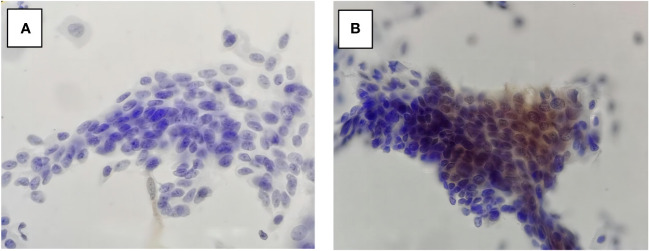
Few articles have focused on the cytological misinterpretation of high-grade squamous intraepithelial lesion (HSIL). Due to estrogen deficiency, cervical epithelial cells in postmenopausal women tend to show atrophic change that looks like HSIL on Papanicolaou-stained cytology slides, resulting in a higher rate of cytological misinterpretation. P16 immunocytochemical staining (P16 cytology) can effectively differentiate diseased cells from normal atrophic ones with less dependence on cell morphology.

OBJECTIVE

To evaluate the role of P16 cytology in differentiating cytology HSIL from benign atrophy in women aged 50 years and above.

METHODS

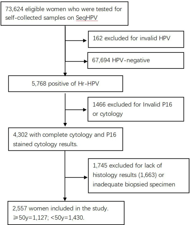
Included in this analysis were women in a cervical cancer screening project conducted in central China who tested positive for high-risk human papillomavirus (hr-HPV) and returned back for triage with complete data of primary HPV testing, liquid-based cytology (LBC) analysis, P16 immuno-stained cytology interpretation, and pathology diagnosis. The included patients were grouped by age: ≥50 (1,127 cases) and <50 years (1,430 cases). The accuracy of LBC and P16 cytology in the detection of pathology ≥HSIL was compared between the two groups, and the role of P16 immuno-stain in differentiating benign cervical lesions from cytology ≥HSIL was further analyzed.

RESULTS

One hundred sixty-seven women (14.8%; 167/1,127) in the ≥50 group and 255 (17.8%, 255/1,430) in the <50 group were pathologically diagnosed as HSIL (Path-HSIL). LBC [≥Atypical Squamous Cell Of Undetermined Significance (ASCUS)] and P16 cytology (positive) respectively detected 63.9% (163/255) and 90.2% (230/255) of the Path-≥HSIL cases in the <50 group and 74.3% (124/167) and 93.4% (124/167) of the Path-≥HSIL cases in the ≥50 group. LBC matched with pathology in 105 (41.2%) of the 255 Path-≥HSIL cases in the <50 group and 93 (55.7%) of the 167 Path-≥HSIL cases in the ≥50 group. There were five in the <50 group and 14 in the ≥50 group that were Path-≤LSIL cases, which were interpreted by LBC as HSIL, but negative in P16 cytology.

CONCLUSION

P16 cytology facilitates differentiation of Path-≤LSIL from LBC-≥HSIL for women 50 years of age and above. It can be used in the lower-resource areas, where qualified cytologists are insufficient, as the secondary screening test for women aged ≥50 to avoid unnecessary biopsies and misinterpretation of LBC primary or secondary screening.

摘要

背景

很少有文章关注高级别鳞状上皮内病变(HSIL)的细胞学误诊。由于雌激素缺乏,绝经后女性的宫颈上皮细胞往往会出现萎缩性改变,在巴氏染色的细胞学涂片上看起来像HSIL,导致细胞学误诊率较高。P16免疫细胞化学染色(P16细胞学)可以有效地区分病变细胞与正常萎缩细胞,对细胞形态的依赖性较小。

目的

评估P16细胞学在50岁及以上女性中将细胞学HSIL与良性萎缩相鉴别的作用。

方法

纳入中国中部一项宫颈癌筛查项目中的女性,这些女性高危型人乳头瘤病毒(hr-HPV)检测呈阳性,并返回进行分流,具备初级HPV检测、液基细胞学(LBC)分析、P16免疫染色细胞学解读和病理诊断的完整数据。纳入的患者按年龄分组:≥50岁(1127例)和<50岁(1430例)。比较两组中LBC和P16细胞学检测病理学≥HSIL的准确性,并进一步分析P16免疫染色在将良性宫颈病变与细胞学≥HSIL相鉴别中的作用。

结果

≥50岁组中有167名女性(14.8%;167/1127),<50岁组中有(255)名女性(17.8%,255/1430)经病理诊断为HSIL(病理HSIL)。LBC[≥意义不明确的非典型鳞状细胞(ASCUS)]和P16细胞学(阳性)分别在<50岁组中检测到255例病理≥HSIL病例中的63.9%(163/255)和90.2%(230/255),在≥50岁组中检测到167例病理≥HSIL病例中的74.3%(124/167)和93.4%(124/167)。在<50岁组的255例病理≥HSIL病例中,LBC与病理相符的有105例(41.2%),在≥50岁组的167例病理≥HSIL病例中,LBC与病理相符的有93例(55.7%)。<50岁组中有5例,≥50岁组中有14例为病理≤低级别鳞状上皮内病变(LSIL)病例,LBC将其解读为HSIL,但P16细胞学为阴性。

结论

P16细胞学有助于50岁及以上女性将病理≤LSIL与LBC≥HSIL相鉴别。在合格细胞病理学家不足的资源匮乏地区,它可作为≥50岁女性的二次筛查试验,以避免不必要的活检以及LBC初次或二次筛查的误诊。

https://cdn.ncbi.nlm.nih.gov/pmc/blobs/3bf7/11165024/b3eaa7778263/fonc-14-1332172-g002.jpg
https://cdn.ncbi.nlm.nih.gov/pmc/blobs/3bf7/11165024/dcd5dc34d1ff/fonc-14-1332172-g001.jpg
https://cdn.ncbi.nlm.nih.gov/pmc/blobs/3bf7/11165024/b3eaa7778263/fonc-14-1332172-g002.jpg
https://cdn.ncbi.nlm.nih.gov/pmc/blobs/3bf7/11165024/dcd5dc34d1ff/fonc-14-1332172-g001.jpg
https://cdn.ncbi.nlm.nih.gov/pmc/blobs/3bf7/11165024/b3eaa7778263/fonc-14-1332172-g002.jpg

相似文献

1
Performance of P16 immunocytochemical stain in facilitating cytology interpretation of HSIL for HPV-positive women aged 50 and above.P16免疫细胞化学染色在辅助50岁及以上HPV阳性女性的高级别鳞状上皮内病变细胞学诊断中的应用
Front Oncol. 2024 May 28;14:1332172. doi: 10.3389/fonc.2024.1332172. eCollection 2024.
2
[Clinical value of p16 immunocytochemistry in cervical cancer screening].p16免疫细胞化学在宫颈癌筛查中的临床价值
Zhonghua Fu Chan Ke Za Zhi. 2020 Nov 25;55(11):784-790. doi: 10.3760/cma.j.cn112141-20200520-00428.
3
Evaluating the Performance of p16 Immunocytochemistry in Cervical Cancer Screening.评估p16免疫细胞化学在宫颈癌筛查中的性能。
Cancer Manag Res. 2020 Sep 25;12:9067-9075. doi: 10.2147/CMAR.S273079. eCollection 2020.
4
Role of Cervical Cancer Biomarkers p16 and Ki67 in Abnormal Cervical Cytological Smear.宫颈癌生物标志物p16和Ki67在宫颈细胞学涂片异常中的作用
J Obstet Gynaecol India. 2021 Feb;71(1):72-77. doi: 10.1007/s13224-020-01380-y. Epub 2020 Nov 18.
5
Utility of p16(ink4a) immunocytochemistry in liquid-based cytology specimens from women treated for high-grade squamous intraepithelial lesions.p16(INK4a)免疫细胞化学在接受高级别鳞状上皮内病变治疗的女性液基细胞学标本中的应用。
Acta Cytol. 2007 Jul-Aug;51(4):517-22. doi: 10.1159/000325787.
6
P16/Ki-67 Dual Staining in Positive Human Papillomavirus DNA Testing for Predictive Diagnosis of Abnormal Cervical Lesions in Northeastern Thai Women.P16/Ki-67 双染色在人乳头瘤病毒 DNA 阳性检测中的应用对预测东北泰国女性宫颈病变的意义。
Asian Pac J Cancer Prev. 2022 Oct 1;23(10):3405-3411. doi: 10.31557/APJCP.2022.23.10.3405.
7
p16/ki-67 dual-stain cytology in the triage of ASCUS and LSIL papanicolaou cytology: results from the European equivocal or mildly abnormal Papanicolaou cytology study.p16/ki-67 双染细胞学在 ASCUS 和 LSIL 巴氏细胞学筛查中的应用:来自欧洲意义不明确或轻度异常巴氏细胞学研究的结果。
Cancer Cytopathol. 2011 Jun 25;119(3):158-66. doi: 10.1002/cncy.20140. Epub 2011 Mar 25.
8
Role of p16(INK4a) cytology testing as an adjunct to enhance the diagnostic specificity and accuracy in human papillomavirus-positive women within an organized cervical cancer screening program.在有组织的宫颈癌筛查项目中,p16(INK4a)细胞学检测作为辅助手段以提高人乳头瘤病毒阳性女性诊断特异性和准确性的作用。
Acta Cytol. 2012;56(5):506-14. doi: 10.1159/000338979. Epub 2012 Sep 27.
9
Liquid-based cytology: do ancillary techniques enhance detection of epithelial abnormalities?液基细胞学:辅助技术是否能提高上皮细胞异常的检出率?
Arch Gynecol Obstet. 2018 Jul;298(1):159-169. doi: 10.1007/s00404-018-4763-z. Epub 2018 May 4.
10
Value of P16 expression in the triage of liquid-based cervical cytology with atypical squamous cells of undetermined significance and low-grade squamous intraepithelial lesions.P16 表达在液基细胞学巴氏涂片检查不典型鳞状细胞意义不明确及低级别鳞状上皮内病变中的分流价值。
Chin Med J (Engl). 2011 Aug;124(16):2443-7.

本文引用的文献

1
HPV persistence after cervical surgical excision of high-grade cervical lesions.高级别宫颈病变宫颈手术切除后HPV持续感染情况
Cancer Cytopathol. 2024 May;132(5):268-269. doi: 10.1002/cncy.22760. Epub 2023 Sep 25.
2
Duration of human papillomavirus persistence and its relationship with recurrent cervical dysplasia.人乳头瘤病毒持续时间及其与复发性宫颈发育不良的关系。
Eur J Cancer Prev. 2023 Nov 1;32(6):525-532. doi: 10.1097/CEJ.0000000000000822. Epub 2023 Jul 3.
3
Performance of p16/Ki67 Immunostaining for Triage of Elderly Women with Atypical Squamous Cells of Undetermined Significance.
p16/Ki67免疫染色在老年意义不明确的非典型鳞状细胞女性分流中的应用
J Clin Med. 2023 May 11;12(10):3400. doi: 10.3390/jcm12103400.
4
Lower accuracy of cytological screening for high-grade squamous intraepithelial neoplasia in women over 50 years of age in Japan.日本 50 岁以上女性中,细胞学筛查高级别鳞状上皮内瘤变的准确性降低。
Int J Clin Oncol. 2022 Feb;27(2):427-433. doi: 10.1007/s10147-021-02065-w. Epub 2021 Dec 6.
5
Evaluation of p16 immunocytology and human papillomavirus (HPV) genotyping triage after primary HPV cervical cancer screening on self-samples in China.中国基于自我采样的原发性人乳头瘤病毒(HPV)宫颈癌筛查后p16免疫细胞化学与HPV基因分型分流评估。
Gynecol Oncol. 2021 Aug;162(2):322-330. doi: 10.1016/j.ygyno.2021.05.014. Epub 2021 May 29.
6
Classification of high-grade cervical intraepithelial neoplasia by p16 , Ki-67, HPV E4 and FAM19A4/miR124-2 methylation status demonstrates considerable heterogeneity with potential consequences for management.通过 p16、Ki-67、HPV E4 和 FAM19A4/miR124-2 甲基化状态对高级别宫颈上皮内瘤变进行分类显示出相当大的异质性,这可能对管理产生影响。
Int J Cancer. 2021 Aug 1;149(3):707-716. doi: 10.1002/ijc.33566. Epub 2021 May 11.
7
Evaluation of Cobas HPV and SeqHPV Assays in the Chinese Multicenter Screening Trial.在中国多中心筛查试验中评估 Cobas HPV 和 SeqHPV 检测方法。
J Low Genit Tract Dis. 2021 Jan 1;25(1):22-26. doi: 10.1097/LGT.0000000000000577.
8
[Clinical value of p16 immunocytochemistry in cervical cancer screening].p16免疫细胞化学在宫颈癌筛查中的临床价值
Zhonghua Fu Chan Ke Za Zhi. 2020 Nov 25;55(11):784-790. doi: 10.3760/cma.j.cn112141-20200520-00428.
9
[The value of p16(INK4a) cytology for early diagnosis of cervical cancer].p16(INK4a)细胞学检查在宫颈癌早期诊断中的价值
Zhonghua Bing Li Xue Za Zhi. 2020 Aug 8;49(8):812-815. doi: 10.3760/cma.j.cn112151-20191203-00779.
10
Pattern of cervical biopsy results in cases with cervical cytology interpreted as higher than low grade in the background with atrophic cellular changes.在存在萎缩性细胞改变背景下,宫颈细胞学检查结果被解读为高于低级别时的宫颈活检结果模式。
Cytojournal. 2020 May 16;17:12. doi: 10.25259/Cytojournal_82_2019. eCollection 2020.