Srebro R
J Physiol. 1985 Mar;360:233-46. doi: 10.1113/jphysiol.1985.sp015614.
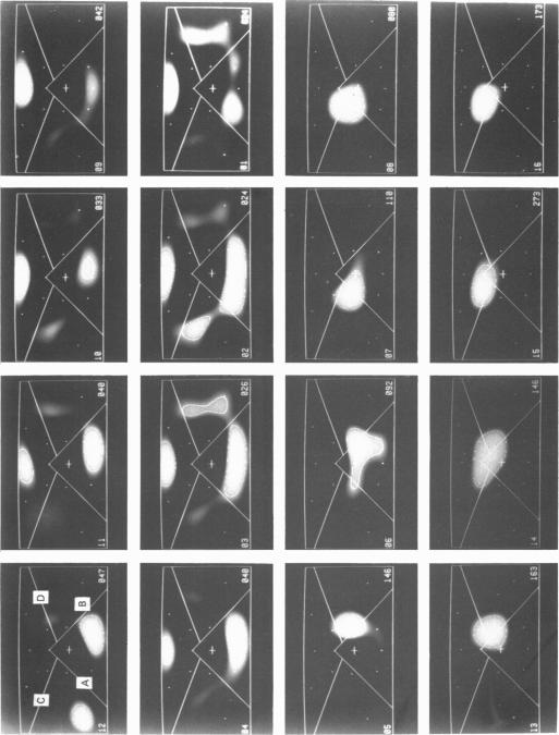
The locations of cortical activity evoked by visual stimuli presented at different positions in the visual field are deduced from the scalp topography of visually evoked potentials in humans. To accomplish this, the Laplacian evoked potential is measured using a multi-electrode array. It is shown that the Laplacian response has the following useful attributes for this purpose. It is reference-free. Its spatial resolution is approximately 2 cm referred to the surface of the cortex. Its spatial sensitivity characteristic is that of a spatial band-pass filter. It is relatively insensitive to source--sink configurations that are oriented tangentially to the surface of the scalp. Only modest assumptions about the source--sink configuration are required to obtain a unique inversion of the scalp topography. Stimuli consisting of checkerboard-filled octant or annular octant segments are presented as appearance-disappearance pulses at sixteen different positions in the visual field in randomized order. The locations of evoked cortical activity in the occipital, parietal and temporal lobes are represented on a Mercator projection map for each octant or octant segment stimulated. Lower hemifield stimuli activate cortex which lies mainly on the convexity of the occipital lobe contralateral to the side of stimulus presentation in the visual field. The more peripheral the stimulus is in the visual field, the more rostral is the location of the active cortex. The rostral-to-caudal location of the evoked activity varies from subject to subject by as much as 3 cm on the surface of the occipital cortex. Furthermore, in any single subject there is a substantial amount of hemispheric asymmetry. Upper hemifield stimuli activate cortex that lies on the extreme caudal pole of the occipital lobe. This activity is relatively weak, and in some subjects it is almost unmeasurable. It is suggested that the representation of the upper hemifield in the cortex lies mostly on the inferior and mesial walls of the occipital lobe and possibly within the calcarine fissures. Those locations are inaccessible to the Laplacian analysis because the current generators therein may be oriented tangentially to the surface of the overlying scalp. Posterior parietal lobe activity and/or inferior temporal lobe activity is frequently evoked. Different subjects have different patterns of evoked activity. Unilateral or bilateral posterior parietal lobe activity is the most common pattern. Unilateral inferior temporal lobe activity is a less common pattern.(ABSTRACT TRUNCATED AT 400 WORDS)
通过人类视觉诱发电位的头皮地形图,可推断出视野中不同位置呈现的视觉刺激所诱发的皮质活动位置。为此,使用多电极阵列测量拉普拉斯诱发电位。结果表明,拉普拉斯反应在此目的下具有以下有用特性。它无需参考电极。其空间分辨率相对于皮质表面约为2厘米。其空间灵敏度特性为空间带通滤波器的特性。它对与头皮表面相切的源 - 汇配置相对不敏感。只需对源 - 汇配置做适度假设,就能对头皮地形图进行唯一反演。由棋盘格填充的八分圆或环形八分圆片段组成的刺激,以出现 - 消失脉冲的形式,在视野中的16个不同位置以随机顺序呈现。针对每个刺激的八分圆或八分圆片段,在墨卡托投影图上表示枕叶、顶叶和颞叶中诱发皮质活动的位置。下半视野刺激激活的皮质主要位于视野中刺激呈现侧对侧枕叶的凸面上。刺激在视野中越靠外周,活跃皮质的位置越靠前。在枕叶皮质表面,诱发活动从嘴侧到尾侧的位置在不同个体间相差可达3厘米。此外,在任何单个个体中都存在大量的半球不对称性。上半视野刺激激活位于枕叶极尾端的皮质。这种活动相对较弱,在某些个体中几乎无法测量。提示皮质中上半视野的表征大多位于枕叶的下壁和内侧壁,可能在距状裂内。由于其中的电流发生器可能与覆盖头皮的表面相切,这些位置无法用拉普拉斯分析检测到。后顶叶活动和/或颞下叶活动经常被诱发。不同个体有不同的诱发活动模式。单侧或双侧后顶叶活动是最常见的模式。单侧颞下叶活动是较不常见的模式。(摘要截取自400字)