National Clinical Research Center for Ocular Diseases, Eye Hospital, Wenzhou Medical University, Wenzhou, China.
Eye Hospital of Wenzhou Medical University at Hangzhou, Hangzhou, China.
Transl Vis Sci Technol. 2024 Jun 3;13(6):6. doi: 10.1167/tvst.13.6.6.
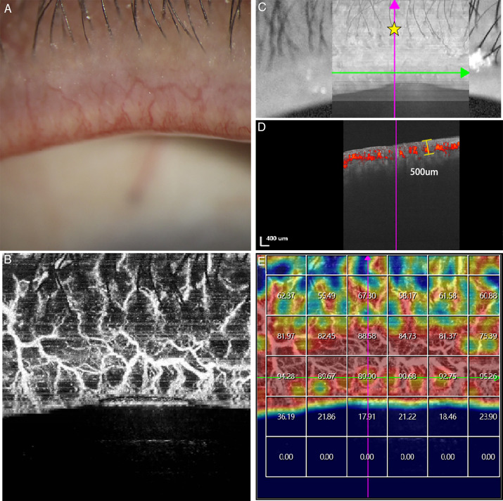
To evaluate the ability of swept-source optical coherence tomography angiography (SS-OCTA) to assess lid margin vascularity.
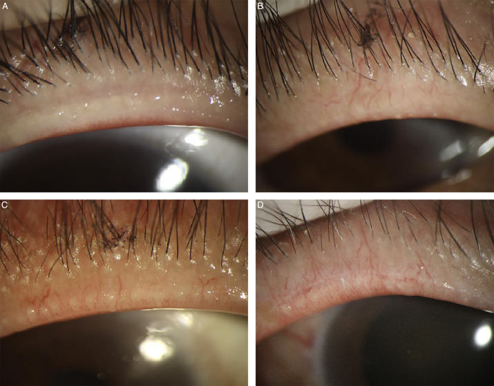
This prospective, cross-sectional trial enrolled 125 participants, including 15 control subjects and 110 meibomian gland dysfunction (MGD) patients. Lid margin blood flow density (LMBFD) was obtained using SS-OCTA. LMBFD was assessed for repeatability in 54 of 125 participants and for reproducibility in 23 of 125 participants. The efficacy of LMBFD was validated in the 125 participants, who were divided into mild (n = 46), moderate (n = 42), and severe groups (n = 37) according to the lid margin vascularity severity shown in the slit-lamp photographs. Correlations between LMBFD and MG-related parameters, such as ocular surface disease index (OSDI), fluorescein tear break-up time (FTBUT), cornea fluorescein staining (CFS), lid margin score (LMS), and meibomian gland expressibility (ME), were analyzed in all 125 participants.
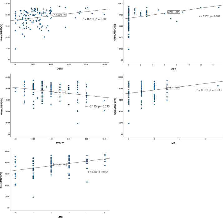
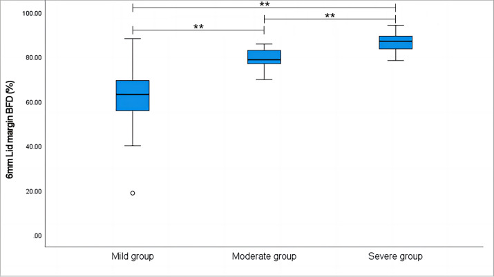
Repeatability and reproducibility coefficients were satisfactorily high in the scan mode with a scan area of 6 mm × 6 mm (intraclass correlation coefficient [ICC] repeatability = 0.905; ICC reproducibility = 0.986) and a scan area of 9 mm × 9 mm (ICC repeatability = 0.888; ICC reproducibility = 0.988). The LMBFD gradually increased in the mild, moderate, and severe groups (P < 0.001). LMBFD was significant correlated with OSDI (r = 0.290, P = 0.001), FTBUT (r = -0.195, P = 0.030), CFS (r = 0.352, P < 0.001), ME (r = 0.191, P = 0.033), and LMS (r = 0.370, P < 0.001).
LMBFD may be a noninvasive, repeatable, reproducible, and efficient index for the quantitative evaluation of eyelid margin vascularity in the future.
We demonstrated that SS-OCTA has the potential to evaluate the eyelid margin vascularity in MGD patients and guide future treatment strategies in clinics.
评估扫频源光相干断层扫描血管造影(SS-OCTA)评估睑缘血管的能力。
本前瞻性、横断面研究纳入了 125 名参与者,包括 15 名对照者和 110 名睑板腺功能障碍(MGD)患者。使用 SS-OCTA 获得睑缘血流密度(LMBFD)。在 125 名参与者中,54 名参与者评估了 LMBFD 的重复性,23 名参与者评估了 LMBFD 的再现性。根据裂隙灯照片显示的睑缘血管严重程度,将 125 名参与者分为轻度(n = 46)、中度(n = 42)和重度组(n = 37),验证 LMBFD 的有效性。分析了所有 125 名参与者中 LMBFD 与眼表疾病指数(OSDI)、泪膜破裂时间(FTBUT)、角膜荧光素染色(CFS)、睑缘评分(LMS)和睑板腺分泌功能(ME)等 MG 相关参数之间的相关性。
使用面积为 6mm×6mm 的扫描模式(组内相关系数[ICC]重复性=0.905;ICC 再现性=0.986)和面积为 9mm×9mm 的扫描模式(ICC 重复性=0.888;ICC 再现性=0.988)时,重复性和再现性系数均较高。LMBFD 在轻度、中度和重度组中逐渐增加(P<0.001)。LMBFD 与 OSDI(r=0.290,P=0.001)、FTBUT(r=-0.195,P=0.030)、CFS(r=0.352,P<0.001)、ME(r=0.191,P=0.033)和 LMS(r=0.370,P<0.001)显著相关。
LMBFD 可能成为未来定量评估睑缘血管的一种非侵入性、可重复、可再现且有效的指标。
杨杰