Department of Biomedical Sciences, University of North Dakota, Grand Forks, ND, USA.
Department of Biomedical Sciences, University of North Dakota, Grand Forks, ND, USA.
J Lipid Res. 2024 Jul;65(7):100583. doi: 10.1016/j.jlr.2024.100583. Epub 2024 Jun 21.
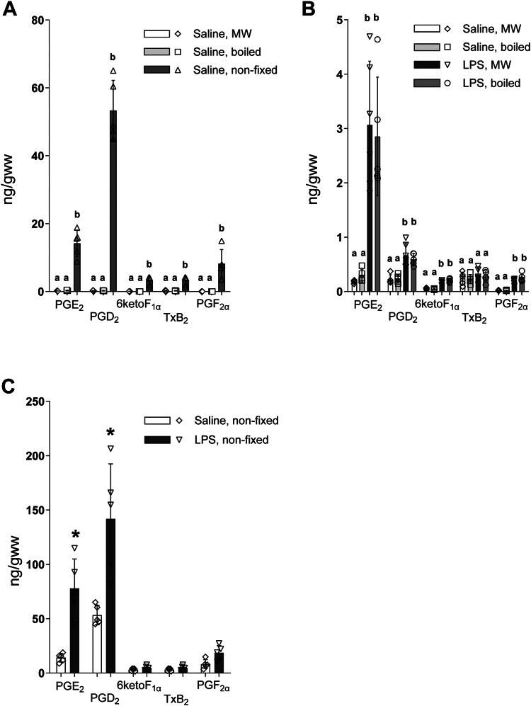
Dramatic postmortem prostanoid (PG) enzymatic synthesis in the brain causes a significant artifact during PG analysis. Thus, enzyme deactivation is required for an accurate in situ endogenous PG quantification. To date, the only method for preventing postmortem brain PG increase with tissue structure preservation is fixation by head-focused microwave irradiation (MW), which is considered the gold standard method, allowing for rapid in situ heat-denaturation of enzymes. However, MW requires costly equipment that suffers in reproducibility, causing tissue loss and metabolite degradation if overheated. Our recent study indicates that PGs are not synthesized in the ischemic brain unless metabolically active tissue is exposed to atmospheric O. Based on this finding, we proposed a simple and reproducible alternative method to prevent postmortem PG increase by slow enzyme denaturation before craniotomy. To test this approach, mice were decapitated directly into boiling saline. Brain temperature reached 100°C after ∼140 s during boiling, though 3 min boiling was required to completely prevent postmortem PG synthesis, but not free arachidonic acid release. To validate this fixation method, brain basal and lipopolysaccharide (LPS)-induced PG were analyzed in unfixed, MW, and boiled tissues. Basal and LPS-induced PG levels were not different between MW and boiled brains. However, unfixed tissue showed a significant postmortem increase in PG at basal conditions, with lesser differences upon LPS treatment compared to fixed tissue. These data indicate for the first time that boiling effectively prevents postmortem PG alterations, allowing for a reproducible, inexpensive, and conventionally accessible tissue fixation method for PG analysis.
在大脑中,剧烈的死后前列腺素(PG)酶促合成会在 PG 分析中产生显著的假象。因此,需要使酶失活,才能准确地进行原位内源性 PG 定量。迄今为止,防止死后大脑 PG 增加并保持组织结构的唯一方法是通过头部聚焦微波辐射(MW)固定,这被认为是金标准方法,可以快速原位热变性酶。然而,MW 需要昂贵的设备,其重复性较差,如果过热,会导致组织损失和代谢物降解。我们最近的研究表明,PG 不会在缺血性大脑中合成,除非代谢活跃的组织暴露于大气 O2 中。基于这一发现,我们提出了一种简单且可重复的替代方法,即在开颅前通过缓慢的酶变性来防止死后 PG 增加。为了验证这种方法,我们将小鼠直接断头浸入沸盐水中。在煮沸过程中,大脑温度在约 140 秒后达到 100°C,尽管需要 3 分钟的煮沸才能完全防止死后 PG 合成,但不会释放游离花生四烯酸。为了验证这种固定方法,我们在未经固定、MW 和煮沸的组织中分析了大脑基础和脂多糖(LPS)诱导的 PG。MW 和煮沸的大脑中的基础和 LPS 诱导的 PG 水平没有差异。然而,未经固定的组织在基础条件下显示出 PG 的显著死后增加,与固定组织相比,LPS 处理后的差异较小。这些数据首次表明,煮沸可有效防止死后 PG 改变,为 PG 分析提供了一种可重复、经济实惠且常规可获得的组织固定方法。