Venianaki Athina P, Barbagianni Mariana S, Fthenakis George C, Galatos Apostolos D, Gouletsou Pagona G
Clinic of Obstetrics and Reproduction, Faculty of Veterinary Science, School of Health Sciences, University of Thessaly, Trikalon 224, 43100 Karditsa, Greece.
Clinic of Surgery, Faculty of Veterinary Science, School of Health Sciences, University of Thessaly, 43100 Karditsa, Greece.
Vet Sci. 2024 Jun 14;11(6):270. doi: 10.3390/vetsci11060270.
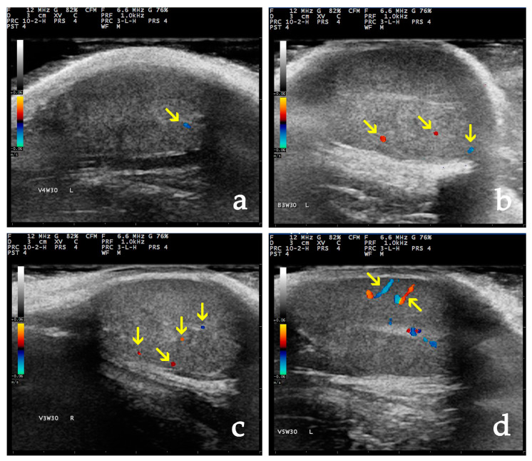
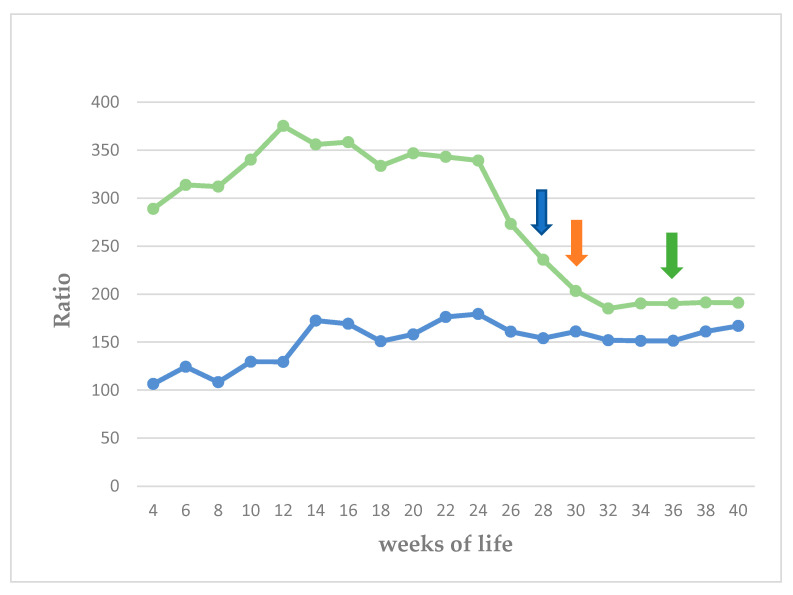
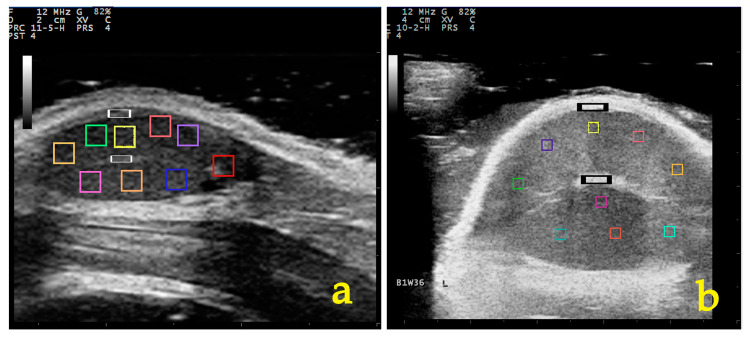
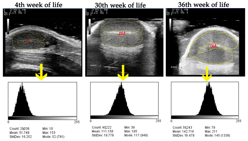
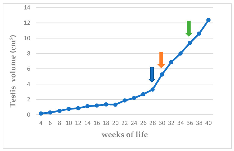
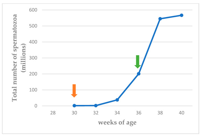
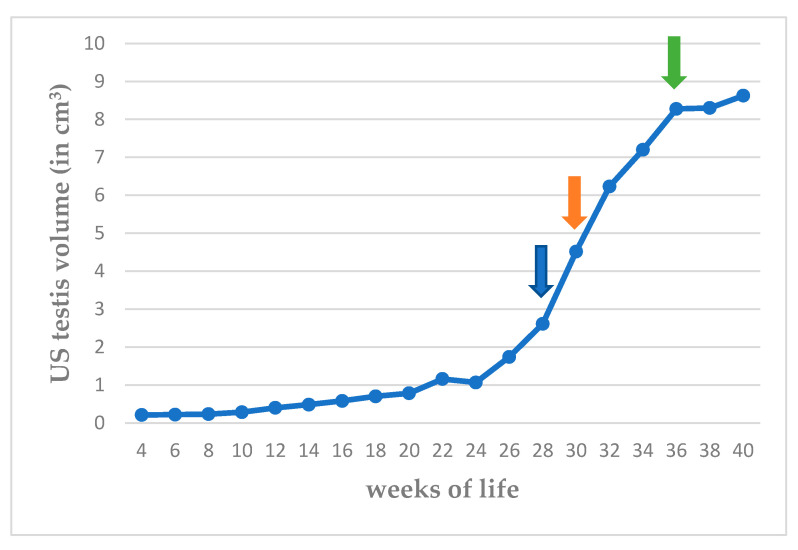
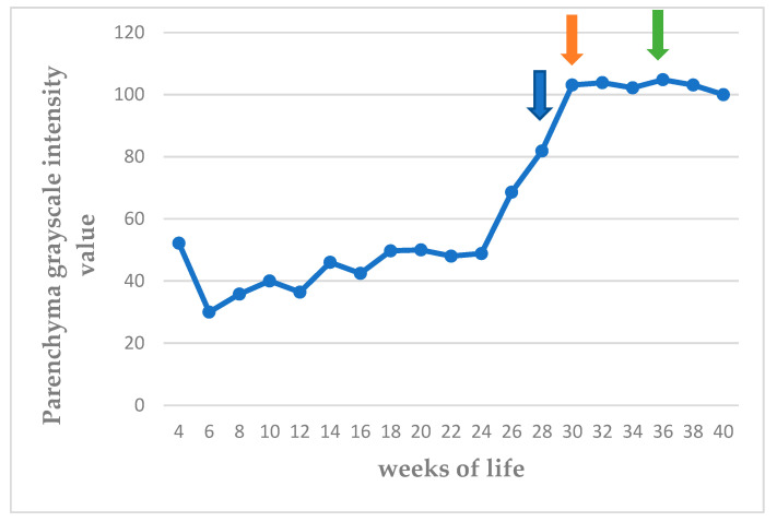
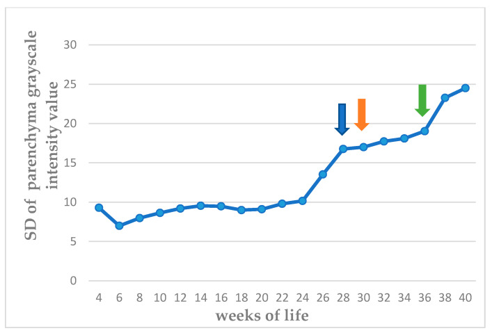
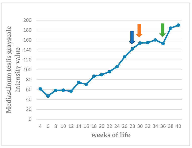
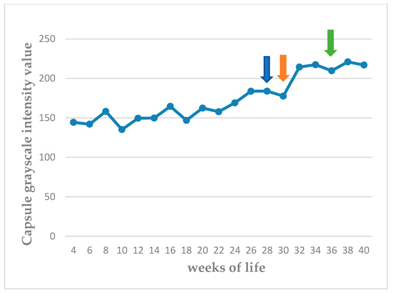
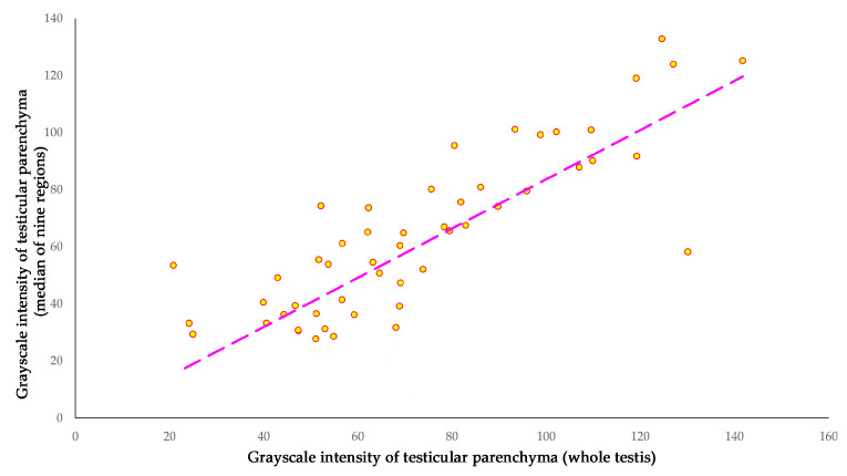
This prospective study investigated the ultrasonographic appearance of the canine testis from birth to adulthood. Eight purpose-bred laboratory Beagle-breed dogs were monitored from 4 to 40 weeks of life. The following parameters were evaluated every two weeks: bodyweight and height, scrotal and testicular volume, ultrasonographically measured testicular volume, echogenicity, heterogeneity, blood-flow score, ratio of the grayscale intensity value of the testis to the capsule, ejaculate volume, motility, viability, and number of spermatozoa. A correlation analysis was carried out between the various measurements obtained. Fertility was achieved in the 36th week of life. The echogenicity of the testicular parenchyma increased with age, and subsequently to the 30th week of life remained constant. The heterogeneity of the testicular parenchyma, as was evaluated by the standard deviation of the values of grayscale intensity of the parenchyma, also increased with age and was >19 at the onset of fertility. The ratio of grayscale intensity of testicular parenchyma had values < 200 at maturity. A colour Doppler evaluation first detected blood flow in the testis in the 22nd week. After the 32nd week, distinct signals were visible. In the 36th week, >80% of the testes imaged had visible vessels. A significant correlation was found between all the evaluation methods. The findings of the study may help clinicians detect the onset of fertility in dogs, especially when semen evaluation is not feasible; however, their applicability in all breeds or individuals might possibly vary due to genetic, physiological, and developmental differences. In summary, the study ultrasonographically explores the testicular maturity in dogs, with the aim to improve clinical assessments and health management in these animals.
这项前瞻性研究调查了犬从出生到成年期睾丸的超声表现。八只专门培育的实验用比格犬从4周龄到40周龄进行监测。每两周评估以下参数:体重和身高、阴囊和睾丸体积、超声测量的睾丸体积、回声性、异质性、血流评分、睾丸与包膜灰度强度值之比、射精量、活力、存活率和精子数量。对获得的各种测量值进行了相关性分析。在第36周龄时达到生育能力。睾丸实质的回声性随年龄增加,随后到第30周龄保持稳定。通过实质灰度强度值的标准差评估的睾丸实质异质性也随年龄增加,在生育开始时>19。睾丸实质灰度强度比在成熟时<200。彩色多普勒评估在第22周首次检测到睾丸内血流。第32周后可见明显信号。在第36周,>80%成像的睾丸有可见血管。所有评估方法之间均发现显著相关性。该研究结果可能有助于临床医生检测犬的生育开始,特别是在精液评估不可行时;然而,由于遗传、生理和发育差异,它们在所有品种或个体中的适用性可能会有所不同。总之,该研究通过超声探索犬的睾丸成熟度,旨在改善这些动物的临床评估和健康管理。