• 文献检索
  • 文档翻译
  • 深度研究
  • 学术资讯
  • Suppr Zotero 插件Zotero 插件
  • 邀请有礼
  • 套餐&价格
  • 历史记录
应用&插件
Suppr Zotero 插件Zotero 插件浏览器插件Mac 客户端Windows 客户端微信小程序
定价
高级版会员购买积分包购买API积分包
服务
文献检索文档翻译深度研究API 文档MCP 服务
关于我们
关于 Suppr公司介绍联系我们用户协议隐私条款
关注我们

Suppr 超能文献

核心技术专利:CN118964589B侵权必究
粤ICP备2023148730 号-1Suppr @ 2026

文献检索

告别复杂PubMed语法,用中文像聊天一样搜索,搜遍4000万医学文献。AI智能推荐,让科研检索更轻松。

立即免费搜索

文件翻译

保留排版,准确专业,支持PDF/Word/PPT等文件格式,支持 12+语言互译。

免费翻译文档

深度研究

AI帮你快速写综述,25分钟生成高质量综述,智能提取关键信息,辅助科研写作。

立即免费体验

超声评估仰卧位胸腔积液量:三种模型公式的比较。

Ultrasound quantification of pleural effusion volume in supine position: comparison of three model formulae.

机构信息

Department of Medical Ultrasound, Tongji Hospital, Tongji Medical College, Huazhong university of Science and Technology, Wuhan, Hubei, 430030, China.

出版信息

BMC Pulm Med. 2024 Jul 4;24(1):316. doi: 10.1186/s12890-024-03142-2.

DOI:10.1186/s12890-024-03142-2
PMID:38965488
原文链接:https://pmc.ncbi.nlm.nih.gov/articles/PMC11225418/
Abstract

BACKGROUND

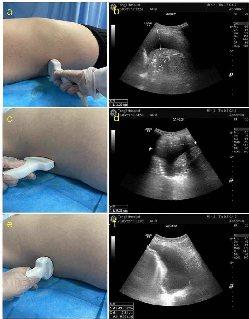
To investigate the accuracy of three model formulae for ultrasound quantification of pleural effusion (PE) volume in patients in supine position.

METHODS

A prospective study including 100 patients with thoracentesis and drainage of PE was conducted. Three model formulae (single section model, two section model and multi-section model) were used to calculate the PE volume. The correlation and consistency analyses between calculated volumes derived from three models and actual PE volume were performed.

RESULTS

PE volumes calculated by three models all showed significant linear correlations with actual PE volume in supine position (all p < 0.001). The reliability of multi-section model in predicting PE volume was significantly higher than that of single section model and slightly higher than that of two section model. When compared with actual drainage volume, the intra-class correlation coefficients (ICCs) of single section model, two section model and multi-section model were 0.72, 0.97 and 0.99, respectively. Significant consistency between calculated PE volumes by using two section model and multi-section model existed for full PE volume range (ICC 0.98).

CONCLUSION

Based on the convenience and accuracy of ultrasound quantification of PE volume, two section model is recommended for pleural effusion assessment in routine clinic, though different model formulae can be selected according to clinical needs.

摘要

背景

探讨三种超声量化平卧位胸腔积液(PE)量的模型公式的准确性。

方法

前瞻性纳入 100 例行胸腔穿刺及引流的 PE 患者。使用三种模型公式(单截面模型、双截面模型和多截面模型)计算 PE 量。分析三种模型计算值与实际 PE 量之间的相关性和一致性。

结果

三种模型计算的 PE 量与平卧位实际 PE 量均呈显著的线性相关(均 p<0.001)。多截面模型预测 PE 量的可靠性明显高于单截面模型,略高于双截面模型。与实际引流量相比,单截面模型、双截面模型和多截面模型的组内相关系数(ICC)分别为 0.72、0.97 和 0.99。双截面模型和多截面模型在全 PE 量范围内计算的 PE 量具有显著的一致性(ICC 0.98)。

结论

基于超声量化 PE 量的便利性和准确性,推荐在常规临床中使用双截面模型评估胸腔积液,但可根据临床需要选择不同的模型公式。

https://cdn.ncbi.nlm.nih.gov/pmc/blobs/3a45/11225418/b3226d391e8a/12890_2024_3142_Fig5_HTML.jpg
https://cdn.ncbi.nlm.nih.gov/pmc/blobs/3a45/11225418/12047b0e452d/12890_2024_3142_Fig1_HTML.jpg
https://cdn.ncbi.nlm.nih.gov/pmc/blobs/3a45/11225418/0db2c6abf6e2/12890_2024_3142_Fig2_HTML.jpg
https://cdn.ncbi.nlm.nih.gov/pmc/blobs/3a45/11225418/55e3b861fb31/12890_2024_3142_Fig3_HTML.jpg
https://cdn.ncbi.nlm.nih.gov/pmc/blobs/3a45/11225418/07a01b411114/12890_2024_3142_Fig4_HTML.jpg
https://cdn.ncbi.nlm.nih.gov/pmc/blobs/3a45/11225418/b3226d391e8a/12890_2024_3142_Fig5_HTML.jpg

相似文献

1
Ultrasound quantification of pleural effusion volume in supine position: comparison of three model formulae.超声评估仰卧位胸腔积液量:三种模型公式的比较。
BMC Pulm Med. 2024 Jul 4;24(1):316. doi: 10.1186/s12890-024-03142-2.
2
Ultrasonographic quantification of pleural effusion: comparison of four formulae.超声对胸腔积液的定量分析:四种公式的比较
Ultrasonography. 2018 Jul;37(3):254-260. doi: 10.14366/usg.17050. Epub 2017 Oct 18.
3
Accuracy of the diagnosis of pleural effusion on supine chest X-ray.仰卧位胸部X线片对胸腔积液诊断的准确性。
Eur Radiol. 1997;7(1):57-60. doi: 10.1007/s003300050109.
4
Multiplane ultrasound approach to quantify pleural effusion at the bedside.床边多平面超声法定量胸腔积液。
Intensive Care Med. 2010 Apr;36(4):656-64. doi: 10.1007/s00134-010-1769-9. Epub 2010 Feb 6.
5
Drainage of pleural effusion in mechanically ventilated patients: time to measure chest wall compliance?机械通气患者胸腔积液的引流:是时候测量胸壁顺应性了吗?
J Crit Care. 2014 Oct;29(5):808-13. doi: 10.1016/j.jcrc.2014.04.009. Epub 2014 Apr 26.
6
CT volumetric analysis of pleural effusions: a comparison with thoracentesis volumes.胸腔积液的CT容积分析:与胸腔穿刺抽液量的比较
Acad Radiol. 2015 Sep;22(9):1122-7. doi: 10.1016/j.acra.2015.03.015. Epub 2015 Jun 22.
7
Quantitative assessment of pleural effusion in critically ill patients by means of ultrasonography.通过超声检查对危重症患者胸腔积液进行定量评估。
Crit Care Med. 2005 Aug;33(8):1757-63. doi: 10.1097/01.ccm.0000171532.02639.08.
8
Ultrasound estimation of volume of pleural fluid in mechanically ventilated patients.机械通气患者胸腔积液量的超声评估
Intensive Care Med. 2006 Feb;32(2):318. doi: 10.1007/s00134-005-0024-2. Epub 2006 Jan 24.
9
Pleural effusions in febrile medical ICU patients: chest ultrasound study.发热的重症医学科患者胸腔积液:胸部超声研究
Chest. 2004 Oct;126(4):1274-80. doi: 10.1378/chest.126.4.1274.
10
Expiratory flow limitation in patients with pleural effusion.胸腔积液患者的呼气气流受限
Respiration. 2007;74(5):572-8. doi: 10.1159/000101489. Epub 2007 Mar 30.

本文引用的文献

1
Automated detection and segmentation of pleural effusion on ultrasound images using an Attention U-net.基于注意力 U-Net 的超声图像胸腔积液自动检测与分割。
J Appl Clin Med Phys. 2024 Jan;25(1):e14231. doi: 10.1002/acm2.14231. Epub 2023 Dec 13.
2
Automatic deep learning-based pleural effusion segmentation in lung ultrasound images.基于深度学习的肺部超声图像中胸腔积液自动分割。
BMC Med Inform Decis Mak. 2023 Nov 29;23(1):274. doi: 10.1186/s12911-023-02362-6.
3
Estimation of Pleural Effusion Volume through Chest Ultrasound: Validation of Two Multiplanar Models.
胸腔超声估算胸腔积液量:两种多平面模型的验证。
Ultrasound Med Biol. 2020 Aug;46(8):1960-1967. doi: 10.1016/j.ultrasmedbio.2020.04.020. Epub 2020 Jun 1.
4
Sonographic Bedside Quantification of Pleural Effusion Compared to Computed Tomography Volumetry in ICU Patients.重症监护病房患者中超声床边定量胸腔积液与计算机断层扫描容积测定法的比较
Ultrasound Int Open. 2018 Oct;4(4):E131-E135. doi: 10.1055/a-0747-6416. Epub 2018 Oct 26.
5
ERS/EACTS statement on the management of malignant pleural effusions.ERS/EACTS 声明:恶性胸腔积液的处理。
Eur J Cardiothorac Surg. 2019 Jan 1;55(1):116-132. doi: 10.1093/ejcts/ezy258.
6
Ultrasonographic quantification of pleural effusion: comparison of four formulae.超声对胸腔积液的定量分析:四种公式的比较
Ultrasonography. 2018 Jul;37(3):254-260. doi: 10.14366/usg.17050. Epub 2017 Oct 18.
7
Validation of equations for pleural effusion volume estimation by ultrasonography.超声检查估计胸腔积液量方程的验证
J Ultrasound. 2017 Oct 27;20(4):267-271. doi: 10.1007/s40477-017-0266-1. eCollection 2017 Dec.
8
Diagnosis and Management of Pleural Transudates.胸腔渗出液的诊断和处理。
Arch Bronconeumol. 2017 Nov;53(11):629-636. doi: 10.1016/j.arbres.2017.04.018. Epub 2017 Jun 19.
9
Computed tomography and patient risk: Facts, perceptions and uncertainties.计算机断层扫描与患者风险:事实、认知与不确定性。
World J Radiol. 2016 Dec 28;8(12):902-915. doi: 10.4329/wjr.v8.i12.902.
10
Nonmalignant Pleural Effusions: A Prospective Study of 356 Consecutive Unselected Patients.非恶性胸腔积液:对356例连续入选的未筛选患者的前瞻性研究。
Chest. 2017 May;151(5):1099-1105. doi: 10.1016/j.chest.2016.12.014. Epub 2016 Dec 23.