• 文献检索
  • 文档翻译
  • 深度研究
  • 学术资讯
  • Suppr Zotero 插件Zotero 插件
  • 邀请有礼
  • 套餐&价格
  • 历史记录
应用&插件
Suppr Zotero 插件Zotero 插件浏览器插件Mac 客户端Windows 客户端微信小程序
定价
高级版会员购买积分包购买API积分包
服务
文献检索文档翻译深度研究API 文档MCP 服务
关于我们
关于 Suppr公司介绍联系我们用户协议隐私条款
关注我们

Suppr 超能文献

核心技术专利:CN118964589B侵权必究
粤ICP备2023148730 号-1Suppr @ 2026

文献检索

告别复杂PubMed语法,用中文像聊天一样搜索,搜遍4000万医学文献。AI智能推荐,让科研检索更轻松。

立即免费搜索

文件翻译

保留排版,准确专业,支持PDF/Word/PPT等文件格式,支持 12+语言互译。

免费翻译文档

深度研究

AI帮你快速写综述,25分钟生成高质量综述,智能提取关键信息,辅助科研写作。

立即免费体验

基于磁共振图像的改进 3D-UNet 脑海马体分割模型。

An improved 3D-UNet-based brain hippocampus segmentation model based on MR images.

机构信息

Information Technology Center, Taizhou University, 1139 Shifu Dadao, Taizhou City, Zhejiang Province, China.

College of Mathematics and Computer Science, Zhejiang A & F University, 666 Wusu Street, Hangzhou, 311300, China.

出版信息

BMC Med Imaging. 2024 Jul 5;24(1):166. doi: 10.1186/s12880-024-01346-w.

DOI:10.1186/s12880-024-01346-w
PMID:38970025
原文链接:https://pmc.ncbi.nlm.nih.gov/articles/PMC11225132/
Abstract

OBJECTIVE

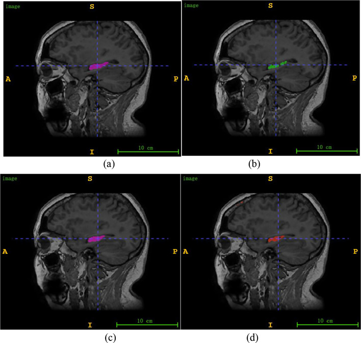
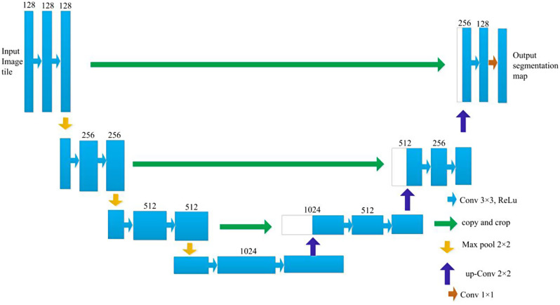
Accurate delineation of the hippocampal region via magnetic resonance imaging (MRI) is crucial for the prevention and early diagnosis of neurosystemic diseases. Determining how to accurately and quickly delineate the hippocampus from MRI results has become a serious issue. In this study, a pixel-level semantic segmentation method using 3D-UNet is proposed to realize the automatic segmentation of the brain hippocampus from MRI results.

METHODS

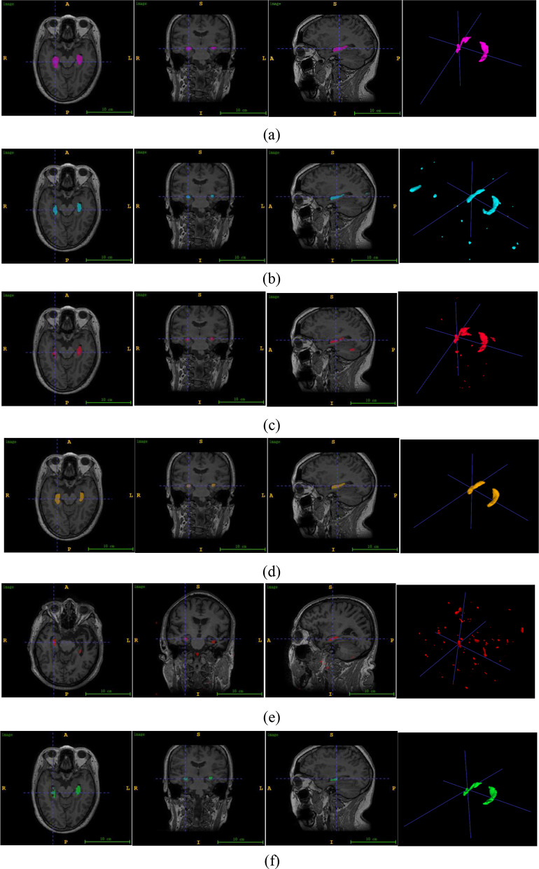
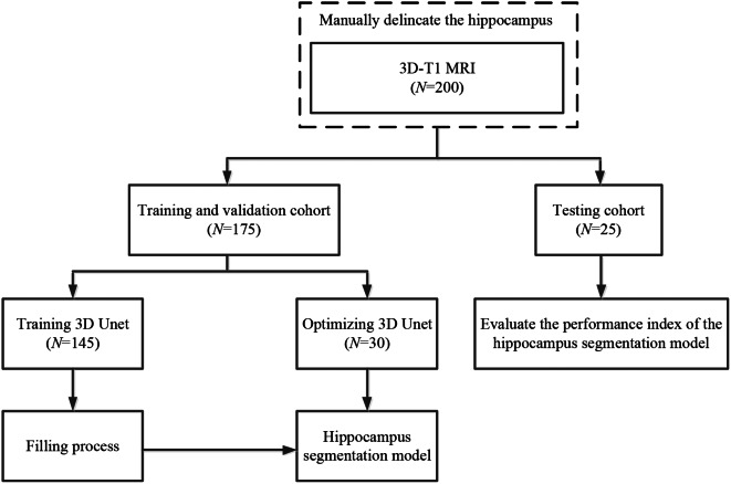
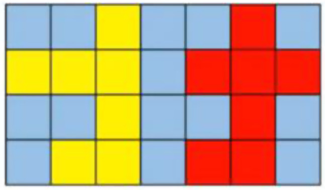
Two hundred three-dimensional T1-weighted (3D-T1) nongadolinium contrast-enhanced magnetic resonance (MR) images were acquired at Hangzhou Cancer Hospital from June 2020 to December 2022. These samples were divided into two groups, containing 175 and 25 samples. In the first group, 145 cases were used to train the hippocampus segmentation model, and the remaining 30 cases were used to fine-tune the hyperparameters of the model. Images for twenty-five patients in the second group were used as the test set to evaluate the performance of the model. The training set of images was processed via rotation, scaling, grey value augmentation and transformation with a smooth dense deformation field for both image data and ground truth labels. A filling technique was introduced into the segmentation network to establish the hippocampus segmentation model. In addition, the performance of models established with the original network, such as VNet, SegResNet, UNetR and 3D-UNet, was compared with that of models constructed by combining the filling technique with the original segmentation network.

RESULTS

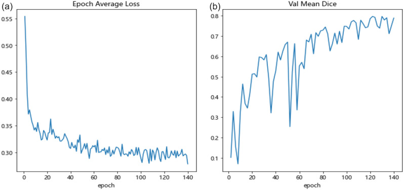
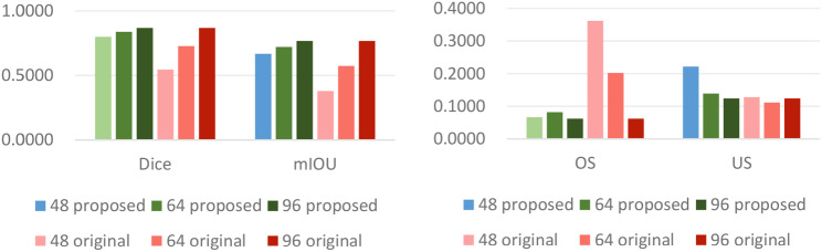
The results showed that the performance of the segmentation model improved after the filling technique was introduced. Specifically, when the filling technique was introduced into VNet, SegResNet, 3D-UNet and UNetR, the segmentation performance of the models trained with an input image size of 48 × 48 × 48 improved. Among them, the 3D-UNet-based model with the filling technique achieved the best performance, with a Dice score (Dice score) of 0.7989 ± 0.0398 and a mean intersection over union (mIoU) of 0.6669 ± 0.0540, which were greater than those of the original 3D-UNet-based model. In addition, the oversegmentation ratio (OSR), average surface distance (ASD) and Hausdorff distance (HD) were 0.0666 ± 0.0351, 0.5733 ± 0.1018 and 5.1235 ± 1.4397, respectively, which were better than those of the other models. In addition, when the size of the input image was set to 48 × 48 × 48, 64 × 64 × 64 and 96 × 96 × 96, the model performance gradually improved, and the Dice scores of the proposed model reached 0.7989 ± 0.0398, 0.8371 ± 0.0254 and 0.8674 ± 0.0257, respectively. In addition, the mIoUs reached 0.6669 ± 0.0540, 0.7207 ± 0.0370 and 0.7668 ± 0.0392, respectively.

CONCLUSION

The proposed hippocampus segmentation model constructed by introducing the filling technique into a segmentation network performed better than models built solely on the original network and can improve the efficiency of diagnostic analysis.

摘要

目的

通过磁共振成像(MRI)准确描绘海马区对于预防和早期诊断神经系统疾病至关重要。如何准确快速地从 MRI 结果中描绘出海马已成为一个严重的问题。本研究提出了一种基于 3D-UNet 的像素级语义分割方法,用于实现从 MRI 结果中自动分割脑海马。

方法

从 2020 年 6 月至 2022 年 12 月,杭州肿瘤医院共采集了 203 例三维 T1 加权(3D-T1)非钆增强磁共振(MR)图像。这些样本分为两组,每组包含 175 例和 25 例。在第一组中,145 例用于训练海马分割模型,其余 30 例用于微调模型的超参数。第二组的 25 例患者的图像用于测试集,以评估模型的性能。对图像训练集进行旋转、缩放、灰度值增强和基于平滑密集变形场的转换处理,同时对图像数据和地面真实标签进行处理。引入填充技术建立海马分割模型。此外,还比较了原始网络(如 VNet、SegResNet、UNetR 和 3D-UNet)建立的模型的性能与原始分割网络与填充技术相结合建立的模型的性能。

结果

结果表明,引入填充技术后分割模型的性能得到了提高。具体来说,当在 VNet、SegResNet、3D-UNet 和 UNetR 中引入填充技术时,输入图像大小为 48×48×48 的模型训练性能得到了提高。其中,基于 3D-UNet 的填充技术模型表现最佳,其 Dice 得分(Dice score)为 0.7989±0.0398,平均交集(mean intersection over union,mIoU)为 0.6669±0.0540,优于原始 3D-UNet 模型。此外,过分割比(oversegmentation ratio,OSR)、平均表面距离(average surface distance,ASD)和 Hausdorff 距离(Hausdorff distance,HD)分别为 0.0666±0.0351、0.5733±0.1018 和 5.1235±1.4397,优于其他模型。此外,当输入图像大小设置为 48×48×48、64×64×64 和 96×96×96 时,模型性能逐渐提高,所提出模型的 Dice 得分分别达到 0.7989±0.0398、0.8371±0.0254 和 0.8674±0.0257,mIoU 分别达到 0.6669±0.0540、0.7207±0.0370 和 0.7668±0.0392。

结论

本研究提出的通过在分割网络中引入填充技术构建的海马分割模型优于仅基于原始网络构建的模型,能够提高诊断分析的效率。

https://cdn.ncbi.nlm.nih.gov/pmc/blobs/1c36/11225132/0e71925efbe3/12880_2024_1346_Fig7_HTML.jpg
https://cdn.ncbi.nlm.nih.gov/pmc/blobs/1c36/11225132/551b3245e7a9/12880_2024_1346_Fig1_HTML.jpg
https://cdn.ncbi.nlm.nih.gov/pmc/blobs/1c36/11225132/d285a7ba233d/12880_2024_1346_Fig2_HTML.jpg
https://cdn.ncbi.nlm.nih.gov/pmc/blobs/1c36/11225132/837f11f81e6b/12880_2024_1346_Fig3_HTML.jpg
https://cdn.ncbi.nlm.nih.gov/pmc/blobs/1c36/11225132/515df92156a1/12880_2024_1346_Fig4_HTML.jpg
https://cdn.ncbi.nlm.nih.gov/pmc/blobs/1c36/11225132/effb5aadca71/12880_2024_1346_Fig5_HTML.jpg
https://cdn.ncbi.nlm.nih.gov/pmc/blobs/1c36/11225132/ecc5207a3678/12880_2024_1346_Fig6_HTML.jpg
https://cdn.ncbi.nlm.nih.gov/pmc/blobs/1c36/11225132/0e71925efbe3/12880_2024_1346_Fig7_HTML.jpg

相似文献

1
An improved 3D-UNet-based brain hippocampus segmentation model based on MR images.基于磁共振图像的改进 3D-UNet 脑海马体分割模型。
BMC Med Imaging. 2024 Jul 5;24(1):166. doi: 10.1186/s12880-024-01346-w.
2
[Fully Automatic Glioma Segmentation Algorithm of Magnetic Resonance Imaging Based on 3D-UNet With More Global Contextual Feature Extraction: An Improvement on Insufficient Extraction of Global Features].基于具有更多全局上下文特征提取的3D-UNet的磁共振成像全自动胶质瘤分割算法:对全局特征提取不足的改进
Sichuan Da Xue Xue Bao Yi Xue Ban. 2024 Mar 20;55(2):447-454. doi: 10.12182/20240360208.
3
Deep learning-based recognition and segmentation of intracranial aneurysms under small sample size.小样本量下基于深度学习的颅内动脉瘤识别与分割
Front Physiol. 2022 Dec 19;13:1084202. doi: 10.3389/fphys.2022.1084202. eCollection 2022.
4
A benchmark study of convolutional neural networks in fully automatic segmentation of aortic root.卷积神经网络在主动脉根部全自动分割中的基准研究。
Front Bioeng Biotechnol. 2023 Jun 15;11:1171868. doi: 10.3389/fbioe.2023.1171868. eCollection 2023.
5
Label-driven magnetic resonance imaging (MRI)-transrectal ultrasound (TRUS) registration using weakly supervised learning for MRI-guided prostate radiotherapy.基于弱监督学习的标签驱动 MRI-经直肠超声(TRUS)配准在 MRI 引导前列腺放疗中的应用。
Phys Med Biol. 2020 Jun 26;65(13):135002. doi: 10.1088/1361-6560/ab8cd6.
6
Computerized Characterization of Spinal Structures on MRI and Clinical Significance of 3D Reconstruction of Lumbosacral Intervertebral Foramen.基于 MRI 的脊柱结构计算机特征化与腰骶椎间孔三维重建的临床意义。
Pain Physician. 2022 Jan;25(1):E27-E35.
7
Automatic intraprostatic lesion segmentation in multiparametric magnetic resonance images with proposed multiple branch UNet.利用提出的多分支U-Net在多参数磁共振图像中实现前列腺内病变的自动分割。
Med Phys. 2020 Dec;47(12):6421-6429. doi: 10.1002/mp.14517. Epub 2020 Oct 24.
8
SK-Unet++: An improved Unet++ network with adaptive receptive fields for automatic segmentation of ultrasound thyroid nodule images.SK-Unet++:一种具有自适应感受野的改进型Unet++网络,用于超声甲状腺结节图像的自动分割。
Med Phys. 2024 Mar;51(3):1798-1811. doi: 10.1002/mp.16672. Epub 2023 Aug 22.
9
Fully automated cardiac MRI segmentation using dilated residual network.使用扩张残差网络的全自动心脏磁共振成像分割
Med Phys. 2023 Apr;50(4):2162-2175. doi: 10.1002/mp.16108. Epub 2022 Dec 7.
10
MI-UNet: Multi-Inputs UNet Incorporating Brain Parcellation for Stroke Lesion Segmentation From T1-Weighted Magnetic Resonance Images.MI-UNet:一种多输入 U 型网络,结合脑区划分用于从 T1 加权磁共振图像中分割中风病灶。
IEEE J Biomed Health Inform. 2021 Feb;25(2):526-535. doi: 10.1109/JBHI.2020.2996783. Epub 2021 Feb 5.

本文引用的文献

1
Plant leaf infected spot segmentation using robust encoder-decoder cascaded deep learning model.基于鲁棒编码器-解码器级联深度学习模型的植物叶片感染斑点分割
Network. 2023 Nov 21:1-19. doi: 10.1080/0954898X.2023.2286002.
2
Bone tumor examination based on FCNN-4s and CRF fine segmentation fusion algorithm.基于全卷积神经网络-4s(FCNN-4s)和条件随机场(CRF)精细分割融合算法的骨肿瘤检测
J Bone Oncol. 2023 Sep 6;42:100502. doi: 10.1016/j.jbo.2023.100502. eCollection 2023 Oct.
3
Intra-oral scan segmentation using deep learning.口腔内扫描分割的深度学习方法。
BMC Oral Health. 2023 Sep 5;23(1):643. doi: 10.1186/s12903-023-03362-8.
4
Active bone marrow segmentation based on computed tomography imaging in anal cancer patients: A machine-learning-based proof of concept.基于 CT 成像的直肠癌患者活跃骨髓自动分割:基于机器学习的概念验证。
Phys Med. 2023 Sep;113:102657. doi: 10.1016/j.ejmp.2023.102657. Epub 2023 Aug 9.
5
Tooth automatic segmentation from CBCT images: a systematic review.基于锥形束计算机断层扫描(CBCT)图像的牙齿自动分割:一项系统综述
Clin Oral Investig. 2023 Jul;27(7):3363-3378. doi: 10.1007/s00784-023-05048-5. Epub 2023 May 6.
6
Agreement Between Automated and Manual MRI Volumetry in Alzheimer's Disease: A Systematic Review and Meta-Analysis.阿尔茨海默病的自动与手动 MRI 容积测量一致性:系统评价和荟萃分析。
J Magn Reson Imaging. 2022 Aug;56(2):490-507. doi: 10.1002/jmri.28037. Epub 2021 Dec 29.
7
Orchard Mapping with Deep Learning Semantic Segmentation.深度学习语义分割的果园图绘制。
Sensors (Basel). 2021 May 31;21(11):3813. doi: 10.3390/s21113813.
8
Liver tumor segmentation using 2.5D UV-Net with multi-scale convolution.使用多尺度卷积的 2.5D UV-Net 进行肝脏肿瘤分割。
Comput Biol Med. 2021 Jun;133:104424. doi: 10.1016/j.compbiomed.2021.104424. Epub 2021 Apr 24.
9
Deep learning-based automatic delineation of the hippocampus by MRI: geometric and dosimetric evaluation.基于深度学习的 MRI 自动勾画海马:几何和剂量评估。
Radiat Oncol. 2021 Jan 14;16(1):12. doi: 10.1186/s13014-020-01724-y.
10
Hippocampus segmentation in CT using deep learning: impact of MR versus CT-based training contours.基于深度学习的CT图像中海马体分割:基于磁共振成像(MR)与基于CT的训练轮廓的影响
J Med Imaging (Bellingham). 2020 Nov;7(6):064001. doi: 10.1117/1.JMI.7.6.064001. Epub 2020 Nov 11.