Department of Neuroradiology, Heidelberg University Hospital, Im Neuenheimer Feld 400, 69120, Heidelberg, Germany.
Clinical Cooperation Unit Neuroimmunology and Brain Tumor Immunology, German Cancer Consortium (DTK) within the German Cancer Research Center (DKFZ), Heidelberg, Germany.
Sci Rep. 2024 Jul 6;14(1):15613. doi: 10.1038/s41598-024-66519-7.
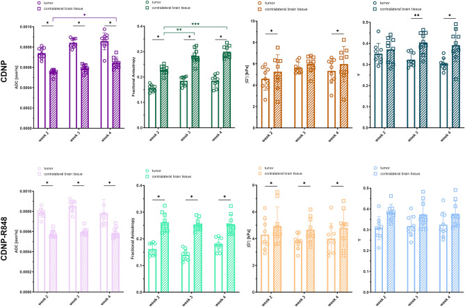
Glioblastoma is the most common and aggressive primary malignant brain tumor with poor prognosis. Novel immunotherapeutic approaches are currently under investigation. Even though magnetic resonance imaging (MRI) is the most important imaging tool for treatment monitoring, response assessment is often hampered by therapy-related tissue changes. As tumor and therapy-associated tissue reactions differ structurally, we hypothesize that biomechanics could be a pertinent imaging proxy for differentiation. Longitudinal MRI and magnetic resonance elastography (MRE) were performed to monitor response to immunotherapy with a toll-like receptor 7/8 agonist in orthotopic syngeneic experimental glioma. Imaging results were correlated to histology and light sheet microscopy data. Here, we identify MRE as a promising non-invasive imaging method for immunotherapy-monitoring by quantifying changes in response-related tumor mechanics. Specifically, we show that a relative softening of treated compared to untreated tumors is linked to the inflammatory processes following therapy-induced re-education of tumor-associated myeloid cells. Mechanistically, combined effects of myeloid influx and inflammation including extracellular matrix degradation following immunotherapy form the basis of treated tumors being softer than untreated glioma. This is a very early indicator of therapy response outperforming established imaging metrics such as tumor volume. The overall anti-tumor inflammatory processes likely have similar effects on human brain tissue biomechanics, making MRE a promising tool for gauging response to immunotherapy in glioma patients early, thereby strongly impacting patient pathway.
胶质母细胞瘤是最常见和侵袭性最强的原发性恶性脑肿瘤,预后不良。目前正在研究新的免疫治疗方法。尽管磁共振成像(MRI)是治疗监测中最重要的成像工具,但反应评估常常受到治疗相关组织变化的阻碍。由于肿瘤和治疗相关的组织反应在结构上不同,我们假设生物力学可能是一种相关的成像替代物。进行了纵向 MRI 和磁共振弹性成像(MRE)以监测 Toll 样受体 7/8 激动剂在原位同基因实验性脑胶质瘤中的免疫治疗反应。将成像结果与组织学和光片显微镜数据相关联。在这里,我们通过量化与治疗相关的肿瘤力学变化,确定 MRE 是一种有前途的免疫治疗监测的非侵入性成像方法。具体来说,我们表明,与未经治疗的肿瘤相比,治疗后的肿瘤相对软化与治疗诱导的肿瘤相关髓样细胞再教育后的炎症过程有关。从机制上讲,髓样细胞浸润和炎症的综合作用,包括免疫治疗后细胞外基质的降解,是治疗后的肿瘤比未经治疗的神经胶质瘤更软的基础。这是治疗反应的一个非常早期的指标,优于肿瘤体积等既定的成像指标。总体抗肿瘤炎症过程可能对人类脑组织生物力学有类似的影响,使 MRE 成为早期评估胶质母细胞瘤患者免疫治疗反应的有前途的工具,从而对患者的治疗路径产生重大影响。