Yu Tianyi, Yan Zirong, Li Zixiang, Yang Meng, Yu Zesen, Chen Yuanjie, Li Wang
Department of Urology, The Affiliated Hospital of Xuzhou Medical University, Xuzhou, China.
Transl Androl Urol. 2024 Jun 30;13(6):949-961. doi: 10.21037/tau-23-656. Epub 2024 Jun 27.
There is lack of discrimination as to traditional imaging diagnostic methods of cystic renal lesions (CRLs). This study aimed to evaluate the value of machine learning models based on clinical data and contrast-enhanced computed tomography (CECT) radiomics features in the differential diagnosis of benign and malignant CRL.
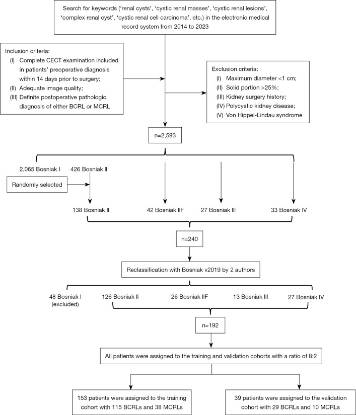
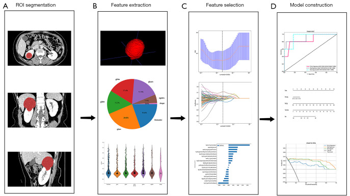
There were 192 patients with CRL (Bosniak class ≥ II) enrolled through histopathological examination, including 144 benign cystic renal lesions (BCRLs) and 48 malignant cystic renal lesions (MCRLs). Radiomics features were extracted from CECT images taken during the medullary phase. Using the light gradient boosting machine (LightGBM) algorithm, the clinical, radiomics and combined models were constructed. A comprehensive nomogram was developed by integrating the radiomics score (Rad-score) with independent clinical factors. Receiver operating characteristic (ROC) curves were plotted. The corresponding area under the curve (AUC) value was worked out to quantify the discrimination performance of the three models in training and validation cohorts. Calibration curves were worked out to assess the accuracy of the probability values predicted by the models. Decision curve analysis (DCA) was worked out to assess the performance of models at different thresholds.
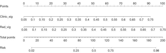
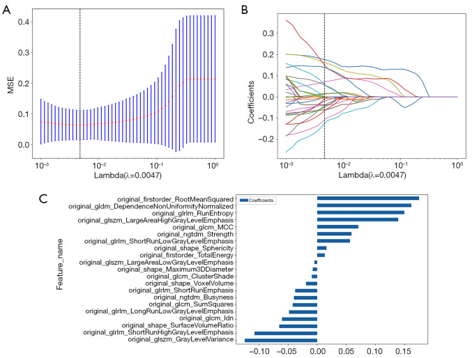
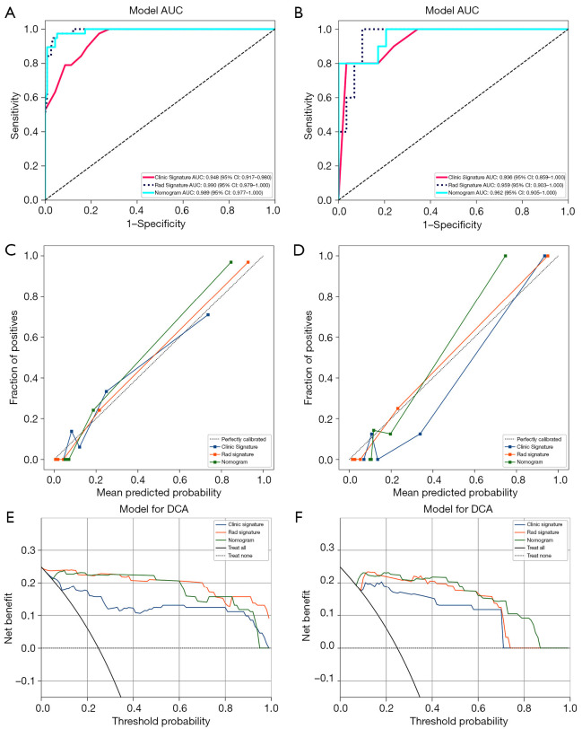
Maximum diameter and Bosniak class were independent risk factors of patients with MCRL in the clinical model. Twenty-one radiomics features were extracted to work out a Rad-score. The performance of the clinical model in the training cohort was AUC =0.948, 95% confidence interval (CI): 0.917-0.980, and the performance in the validation cohort was AUC =0.936, 95% CI: 0.859-1.000 (P<0.05). The performance of the radiomics model in the training cohort was AUC =0.990, 95% CI: 0.979-1.000, and the performance in the validation cohort was AUC =0.959, 95% CI: 0.903-1.000 (P<0.05). Compared with the above models, the combined radiomics nomogram had an AUC of 0.989 (95% CI: 0.977-1.000) in the training cohort and an AUC of 0.962 (95% CI: 0.905-1.000) in the validation cohort (P<0.05), showing the best diagnostic efficacy.
The radiomics nomogram integrating clinical independent risk factors and radiomics signature improved the diagnostic accuracy in differentiating between BCRL and MCRL, which can provide a reference for clinical decision-making and help clinicians develop individualized treatment strategies for patients.
对于囊性肾病变(CRL)的传统影像诊断方法缺乏鉴别能力。本研究旨在评估基于临床数据和对比增强计算机断层扫描(CECT)影像组学特征的机器学习模型在良性和恶性CRL鉴别诊断中的价值。
通过组织病理学检查纳入192例CRL患者(Bosniak分级≥II级),其中包括144例良性囊性肾病变(BCRL)和48例恶性囊性肾病变(MCRL)。从髓质期采集的CECT图像中提取影像组学特征。使用轻梯度提升机(LightGBM)算法构建临床、影像组学和联合模型。通过将影像组学评分(Rad-score)与独立临床因素相结合,开发了一个综合列线图。绘制受试者操作特征(ROC)曲线。计算相应的曲线下面积(AUC)值,以量化三个模型在训练和验证队列中的鉴别性能。绘制校准曲线以评估模型预测概率值的准确性。进行决策曲线分析(DCA)以评估模型在不同阈值下的性能。
在临床模型中,最大直径和Bosniak分级是MCRL患者的独立危险因素。提取了21个影像组学特征以计算Rad-score。临床模型在训练队列中的性能为AUC =0.948,95%置信区间(CI):0.917 - 0.980,在验证队列中的性能为AUC =0.936,95%CI:0.859 - 1.000(P<0.05)。影像组学模型在训练队列中的性能为AUC =0.990,95%CI:0.979 - 1.000,在验证队列中的性能为AUC =0.959,95%CI:0.903 - 1.000(P<0.05)。与上述模型相比,联合影像组学列线图在训练队列中的AUC为0.989(95%CI:0.977 - 1.000),在验证队列中的AUC为0.962(95%CI:0.905 - 1.000)(P<0.05),显示出最佳诊断效能。
整合临床独立危险因素和影像组学特征的影像组学列线图提高了鉴别BCRL和MCRL的诊断准确性,可为临床决策提供参考,并帮助临床医生为患者制定个体化治疗策略。