Department of Ophthalmology, Albert Eye Research Institute, Duke University, Durham, NC 27710, USA.
Department of Ophthalmology, Albert Eye Research Institute, Duke University, Durham, NC 27710, USA.
STAR Protoc. 2024 Sep 20;5(3):103150. doi: 10.1016/j.xpro.2024.103150. Epub 2024 Jul 13.
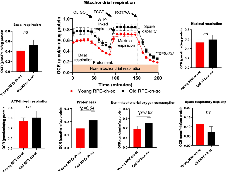
During aging and in retinal degenerative diseases, vulnerable retinal pigment epithelial (RPE) cells are subject to mitochondrial dysfunction, creating a need for accessibility to tools which can facilitate assessment of the ocular posterior pole bioenergetics. Here, we present a protocol for quantifying mitochondrial respiration in the posterior eye cup (RPE-choroid-sclera) of young and old mice. We describe steps for eye cup dissection, optimization of tissue size, drug concentrations, and cycle conditions using the XF Cell Mito Stress Test.
在衰老和视网膜退行性疾病中,脆弱的视网膜色素上皮 (RPE) 细胞易发生线粒体功能障碍,因此需要能够方便地评估眼后极生物能量的工具。在这里,我们提供了一种在年轻和老年小鼠的后眼杯(RPE-脉络膜-巩膜)中定量测量线粒体呼吸的方案。我们描述了使用 XF 细胞线粒体应激测试进行眼杯解剖、组织大小优化、药物浓度和循环条件优化的步骤。