Department of Medical Imaging, Institut de Recherche Expérimentale et Clinique (IREC), Institut du Cancer Roi Albert II, Cliniques Universitaires Saint Luc, Université Catholique de Louvain (UCL), Avenue Hippocrate, 10, B-1200, Brussels, Belgium.
Skeletal Radiol. 2024 Sep;53(9):1815-1831. doi: 10.1007/s00256-024-04723-2. Epub 2024 Jul 15.
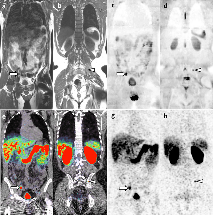
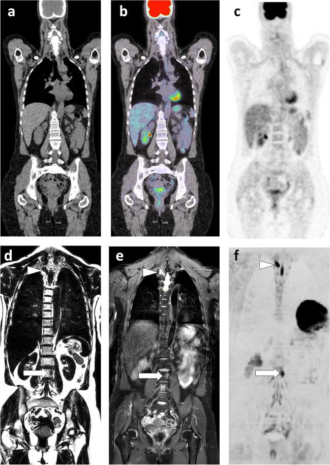
Metastatic disease and myeloma present unique diagnostic challenges due to their multifocal nature. Accurate detection and staging are critical for determining appropriate treatment. Bone scintigraphy, skeletal radiographs and CT have long been the mainstay for the assessment of these diseases, but have limitations, including reduced sensitivity and radiation exposure. Whole-body MRI has emerged as a highly sensitive and radiation-free alternative imaging modality. Initially developed for skeletal screening, it has extended tumor screening to all organs, providing morphological and physiological information on tumor tissue. Along with PET/CT, whole-body MRI is now accepted for staging and response assessment in many malignancies. It is the first choice in an ever increasing number of cancers (such as myeloma, lobular breast cancer, advanced prostate cancer, myxoid liposarcoma, bone sarcoma, …). It has also been validated as the method of choice for cancer screening in patients with a predisposition to cancer and for staging cancers observed during pregnancy. The current and future challenges for WB-MRI are its availability facing this number of indications, and its acceptance by patients, radiologists and health authorities. Guidelines have been developed to optimize image acquisition and reading, assessment of lesion response to treatment, and to adapt examination designs to specific cancers. The implementation of 3D acquisition, Dixon method, and deep learning-based image optimization further improve the diagnostic performance of the technique and reduce examination durations. Whole-body MRI screening is feasible in less than 30 min. This article reviews validated indications, recent developments, growing acceptance, and future perspectives of whole-body MRI.
转移性疾病和骨髓瘤由于其多灶性的性质,给诊断带来了独特的挑战。准确的检测和分期对于确定适当的治疗至关重要。骨闪烁扫描、骨骼 X 线摄影和 CT 长期以来一直是评估这些疾病的主要方法,但存在局限性,包括敏感性降低和辐射暴露。全身 MRI 已成为一种高度敏感且无辐射的替代成像方式。最初开发用于骨骼筛查,现已将肿瘤筛查扩展到所有器官,为肿瘤组织提供形态和生理信息。全身 MRI 与 PET/CT 一起,现在已被许多恶性肿瘤的分期和反应评估所接受。在越来越多的癌症中(如骨髓瘤、乳腺小叶癌、晚期前列腺癌、黏液样脂肪肉瘤、骨肉瘤等),它已成为首选方法。它还已被验证为癌症易感性患者癌症筛查和妊娠期间观察到的癌症分期的首选方法。目前和未来 WB-MRI 的挑战在于它在如此多的适应症中的可用性,以及患者、放射科医生和卫生当局对它的接受程度。已经制定了指南来优化图像采集和阅读、评估病变对治疗的反应,并使检查设计适应特定的癌症。3D 采集、Dixon 方法和基于深度学习的图像优化的应用进一步提高了该技术的诊断性能并缩短了检查时间。全身 MRI 筛查可在 30 分钟内完成。本文回顾了全身 MRI 的验证适应证、最新进展、日益增加的接受度以及未来展望。