• 文献检索
  • 文档翻译
  • 深度研究
  • 学术资讯
  • Suppr Zotero 插件Zotero 插件
  • 邀请有礼
  • 套餐&价格
  • 历史记录
应用&插件
Suppr Zotero 插件Zotero 插件浏览器插件Mac 客户端Windows 客户端微信小程序
定价
高级版会员购买积分包购买API积分包
服务
文献检索文档翻译深度研究API 文档MCP 服务
关于我们
关于 Suppr公司介绍联系我们用户协议隐私条款
关注我们

Suppr 超能文献

核心技术专利:CN118964589B侵权必究
粤ICP备2023148730 号-1Suppr @ 2026

文献检索

告别复杂PubMed语法,用中文像聊天一样搜索,搜遍4000万医学文献。AI智能推荐,让科研检索更轻松。

立即免费搜索

文件翻译

保留排版,准确专业,支持PDF/Word/PPT等文件格式,支持 12+语言互译。

免费翻译文档

深度研究

AI帮你快速写综述,25分钟生成高质量综述,智能提取关键信息,辅助科研写作。

立即免费体验

通过可见光光学相干断层扫描新技术观察到的中央凹高反射点

Hyperreflective Dots in Central Fovea Visualized by a Novel Application of Visible-Light Optical Coherence Tomography.

作者信息

Krause Michael A, Grannonico Marta, Tyler Brooke P, Miller David A, Fan Weijia, Liu Mingna, Kuranov Roman V, Zhang Hao F, Liu Xiaorong, Netland Peter A

机构信息

Department of Ophthalmology University of Virginia, Charlottesville, Virginia, USA.

Department of Biology University of Virginia, Charlottesville, Virginia, USA.

出版信息

Case Rep Ophthalmol Med. 2024 Jul 9;2024:5823455. doi: 10.1155/2024/5823455. eCollection 2024.

DOI:10.1155/2024/5823455
PMID:39015384
原文链接:https://pmc.ncbi.nlm.nih.gov/articles/PMC11251792/
Abstract

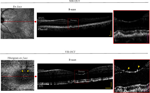
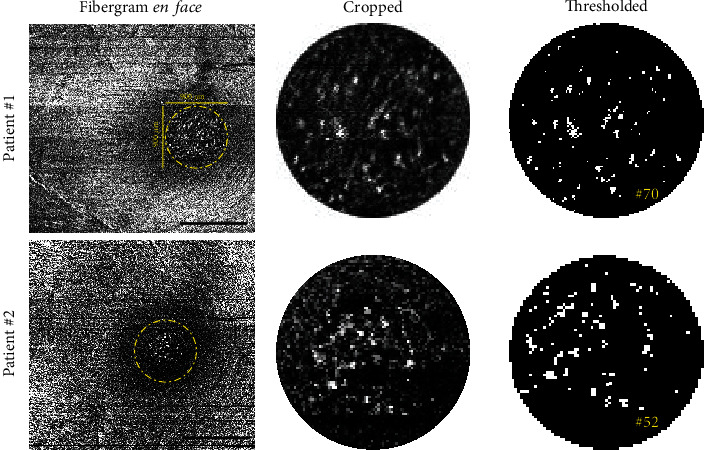
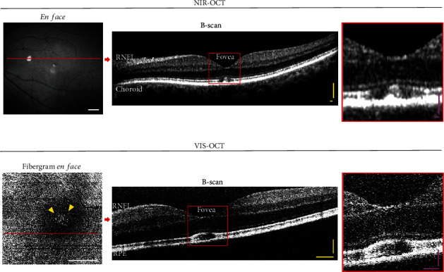
Visible-light optical coherence tomography (vis-OCT) is a novel noninvasive retinal imaging system that offers improved resolution compared to conventional near-infrared (NIR) OCT systems. Here, we utilized vis-OCT to produce fibergrams (vis-OCTF) for the first time in human patients, enabling visualization and precise quantification of hyperreflective dots in the central fovea in two patients. We also directly compare the imaging qualities of conventional vis-OCT and NIR-OCT. Vis-OCT generated a 3 × 3 mm image with an impressive axial resolution of 1.3 m, whereas NIR-OCT produced an image with a larger field of view (FOV) (9 × 9 mm) but a lower resolution of 7.0 m. Moreover, vis-OCTF unveiled clear images of hyperreflective dots in the fovea of both patients, which were not discernible in the NIR-OCT images. Foveal dots have often been linked to several age-related and pathological conditions. The high-resolution images generated by vis-OCTF enable more precise characterization of changes in retinal sublayers within the central fovea.

摘要

可见光光学相干断层扫描(vis-OCT)是一种新型的非侵入性视网膜成像系统,与传统的近红外(NIR)OCT系统相比,它具有更高的分辨率。在此,我们首次在人类患者中利用vis-OCT生成纤维图(vis-OCTF),从而能够可视化并精确量化两名患者中央凹中的高反射点。我们还直接比较了传统vis-OCT和NIR-OCT的成像质量。vis-OCT生成了一幅3×3毫米的图像,其轴向分辨率高达1.3微米,而NIR-OCT生成的图像视野更大(9×9毫米),但分辨率较低,为7.0微米。此外,vis-OCTF揭示了两名患者中央凹中高反射点的清晰图像,而这些在NIR-OCT图像中是无法辨别的。中央凹点常常与多种年龄相关和病理状况有关。vis-OCTF生成的高分辨率图像能够更精确地表征中央凹内视网膜各亚层的变化。

https://cdn.ncbi.nlm.nih.gov/pmc/blobs/dcda/11251792/ba23e167c2ac/CRIOPM2024-5823455.003.jpg
https://cdn.ncbi.nlm.nih.gov/pmc/blobs/dcda/11251792/1a4c31f60cf7/CRIOPM2024-5823455.001.jpg
https://cdn.ncbi.nlm.nih.gov/pmc/blobs/dcda/11251792/40dfa4cd3ac1/CRIOPM2024-5823455.002.jpg
https://cdn.ncbi.nlm.nih.gov/pmc/blobs/dcda/11251792/ba23e167c2ac/CRIOPM2024-5823455.003.jpg
https://cdn.ncbi.nlm.nih.gov/pmc/blobs/dcda/11251792/1a4c31f60cf7/CRIOPM2024-5823455.001.jpg
https://cdn.ncbi.nlm.nih.gov/pmc/blobs/dcda/11251792/40dfa4cd3ac1/CRIOPM2024-5823455.002.jpg
https://cdn.ncbi.nlm.nih.gov/pmc/blobs/dcda/11251792/ba23e167c2ac/CRIOPM2024-5823455.003.jpg

相似文献

1
Hyperreflective Dots in Central Fovea Visualized by a Novel Application of Visible-Light Optical Coherence Tomography.通过可见光光学相干断层扫描新技术观察到的中央凹高反射点
Case Rep Ophthalmol Med. 2024 Jul 9;2024:5823455. doi: 10.1155/2024/5823455. eCollection 2024.
2
Analysis of Hyperreflective Dots Within the Central Fovea in Healthy Eyes Using En Face Optical Coherence Tomography.利用光学相干断层扫描对健康眼中央凹内高反射点的分析。
Invest Ophthalmol Vis Sci. 2019 Oct 1;60(13):4451-4461. doi: 10.1167/iovs.19-27476.
3
Multimodal Imaging, OCT B-Scan Localization, and OCT Detection of Macular Hyperpigmentation in Eyes with Intermediate Age-Related Macular Degeneration.多模态成像、光学相干断层扫描B扫描定位以及光学相干断层扫描检测中度年龄相关性黄斑变性眼中的黄斑色素沉着
Ophthalmol Sci. 2022 Jan 24;2(2):100116. doi: 10.1016/j.xops.2022.100116. eCollection 2022 Jun.
4
En Face Optical Coherence Tomography Imaging of Foveal Dots in Eyes With Posterior Vitreous Detachment or Internal Limiting Membrane Peeling.后玻璃体脱离或内界膜剥除眼的黄斑区小点的光学相干断层扫描成像
Invest Ophthalmol Vis Sci. 2021 Aug 2;62(10):5. doi: 10.1167/iovs.62.10.5.
5
Human retinal imaging using visible-light optical coherence tomography guided by scanning laser ophthalmoscopy.使用扫描激光检眼镜引导的可见光光学相干断层扫描技术进行人体视网膜成像。
Biomed Opt Express. 2015 Sep 1;6(10):3701-13. doi: 10.1364/BOE.6.003701. eCollection 2015 Oct 1.
6
Feasibility of a method for en face imaging of photoreceptor cell integrity.用于检测光感受器细胞完整性的共面成像方法的可行性。
Am J Ophthalmol. 2011 Nov;152(5):807-14.e1. doi: 10.1016/j.ajo.2011.04.027. Epub 2011 Jul 20.
7
Optical coherence tomographic evaluation of foveal hard exudates in patients with diabetic maculopathy accompanying macular detachment.光学相干断层扫描评价伴有黄斑脱离的糖尿病性黄斑病变患者的中心凹硬性渗出物。
Ophthalmology. 2010 Oct;117(10):1996-2002. doi: 10.1016/j.ophtha.2010.06.019. Epub 2010 Aug 17.
8
A Comparison of En Face Optical Coherence Tomography and Fundus Autofluorescence in Stargardt Disease.斯塔加特病中视网膜光学相干断层扫描与眼底自发荧光的比较
Invest Ophthalmol Vis Sci. 2017 Oct 1;58(12):5227-5236. doi: 10.1167/iovs.17-22532.
9
Case report: the first case of unilateral retinal pigment epithelium dysgenesis in China.病例报告:中国首例单侧视网膜色素上皮发育异常病例。
BMC Ophthalmol. 2020 Aug 20;20(1):340. doi: 10.1186/s12886-020-01609-4.
10
Acute Solar Retinopathy Imaged With Adaptive Optics, Optical Coherence Tomography Angiography, and En Face Optical Coherence Tomography.急性太阳视网膜病变的自适应光学成像、光相干断层扫描血管造影和共焦光学相干断层扫描。
JAMA Ophthalmol. 2018 Jan 1;136(1):82-85. doi: 10.1001/jamaophthalmol.2017.5517.

本文引用的文献

1
Visible-Light Optical Coherence Tomography Fibergraphy of the Tree Shrew Retinal Ganglion Cell Axon Bundles.树鼩视网膜神经节细胞轴突束的可见光光学相干断层扫描纤维成像
IEEE Trans Med Imaging. 2024 Aug;43(8):2769-2777. doi: 10.1109/TMI.2024.3380530. Epub 2024 Aug 1.
2
Acute-Onset Retinal Conditions Mimicking Acute Optic Neuritis: Overview and Differential Diagnosis.酷似急性视神经炎的急性发作性视网膜疾病:概述与鉴别诊断
J Clin Med. 2023 Sep 1;12(17):5720. doi: 10.3390/jcm12175720.
3
Longitudinal Analysis of Retinal Ganglion Cell Damage at Individual Axon Bundle Level in Mice Using Visible-Light Optical Coherence Tomography Fibergraphy.
利用可见光光相干断层成像纤维图对小鼠视神经单个轴索束水平的视网膜神经节细胞损伤进行纵向分析。
Transl Vis Sci Technol. 2023 May 1;12(5):10. doi: 10.1167/tvst.12.5.10.
4
In Vivo Sublayer Analysis of Human Retinal Inner Plexiform Layer Obtained by Visible-Light Optical Coherence Tomography.基于可见 光光学相干断层扫描的人视网膜内丛状层的体层分析。
Invest Ophthalmol Vis Sci. 2022 Jan 3;63(1):18. doi: 10.1167/iovs.63.1.18.
5
Low-Reflectivity Drusen With Overlying RPE Damage Revealed by Spectral-Domain OCT: Hint for the Development of Age-Related Macular Degeneration.频域光学相干断层扫描显示的伴有视网膜色素上皮损伤的低反射性玻璃膜疣:年龄相关性黄斑变性发展的线索
Front Med (Lausanne). 2021 Oct 26;8:706502. doi: 10.3389/fmed.2021.706502. eCollection 2021.
6
Visible Light Optical Coherence Tomography (OCT) Quantifies Subcellular Contributions to Outer Retinal Band 4.可见光是相干断层扫描 (OCT) 量化了外视网膜带 4 的亚细胞贡献。
Transl Vis Sci Technol. 2021 Mar 1;10(3):30. doi: 10.1167/tvst.10.3.30.
7
Analysis of Hyperreflective Dots Within the Central Fovea in Healthy Eyes Using En Face Optical Coherence Tomography.利用光学相干断层扫描对健康眼中央凹内高反射点的分析。
Invest Ophthalmol Vis Sci. 2019 Oct 1;60(13):4451-4461. doi: 10.1167/iovs.19-27476.
8
Designing visible-light optical coherence tomography towards clinics.面向临床的可见光光学相干断层扫描技术设计。
Quant Imaging Med Surg. 2019 May;9(5):769-781. doi: 10.21037/qims.2019.05.01.
9
Ultrahigh resolution retinal imaging by visible light OCT with longitudinal achromatization.通过具有纵向消色差功能的可见光光学相干断层扫描实现超高分辨率视网膜成像。
Biomed Opt Express. 2018 Mar 5;9(4):1477-1491. doi: 10.1364/BOE.9.001477. eCollection 2018 Apr 1.
10
Visible-light optical coherence tomography: a review.可见光学相干断层扫描:综述。
J Biomed Opt. 2017 Dec;22(12):1-14. doi: 10.1117/1.JBO.22.12.121707.