• 文献检索
  • 文档翻译
  • 深度研究
  • 学术资讯
  • Suppr Zotero 插件Zotero 插件
  • 邀请有礼
  • 套餐&价格
  • 历史记录
应用&插件
Suppr Zotero 插件Zotero 插件浏览器插件Mac 客户端Windows 客户端微信小程序
定价
高级版会员购买积分包购买API积分包
服务
文献检索文档翻译深度研究API 文档MCP 服务
关于我们
关于 Suppr公司介绍联系我们用户协议隐私条款
关注我们

Suppr 超能文献

核心技术专利:CN118964589B侵权必究
粤ICP备2023148730 号-1Suppr @ 2026

文献检索

告别复杂PubMed语法,用中文像聊天一样搜索,搜遍4000万医学文献。AI智能推荐,让科研检索更轻松。

立即免费搜索

文件翻译

保留排版,准确专业,支持PDF/Word/PPT等文件格式,支持 12+语言互译。

免费翻译文档

深度研究

AI帮你快速写综述,25分钟生成高质量综述,智能提取关键信息,辅助科研写作。

立即免费体验

无需采血的心肌细胞外容积定量分析。

Quantification of myocardial extracellular volume without blood sampling.

作者信息

Chen Wensu, Faragli Alessandro, Goetze Collin, Zieschang Victoria, Weiss Karl Jakob, Hashemi Djawid, Beyer Rebecca, Hafermann Lorena, Stawowy Philipp, Kelle Sebastian, Doeblin Patrick

机构信息

Department of Cardiology, Angiology and Intensive Care Medicine, Deutsches Herzzentrum der Charité, Augustenburger Platz 1, Berlin 13353, Germany.

Charité - Universitätsmedizin Berlin, corporate member of Freie Universität Berlin and Humboldt-Universität zu Berlin, Charitéplatz 1, Berlin 10117, Germany.

出版信息

Eur Heart J Imaging Methods Pract. 2023 Sep 15;1(2):qyad022. doi: 10.1093/ehjimp/qyad022. eCollection 2023 Sep.

DOI:10.1093/ehjimp/qyad022
PMID:39045067
原文链接:https://pmc.ncbi.nlm.nih.gov/articles/PMC11195702/
Abstract

AIMS

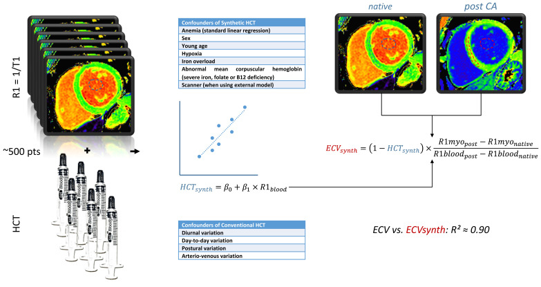
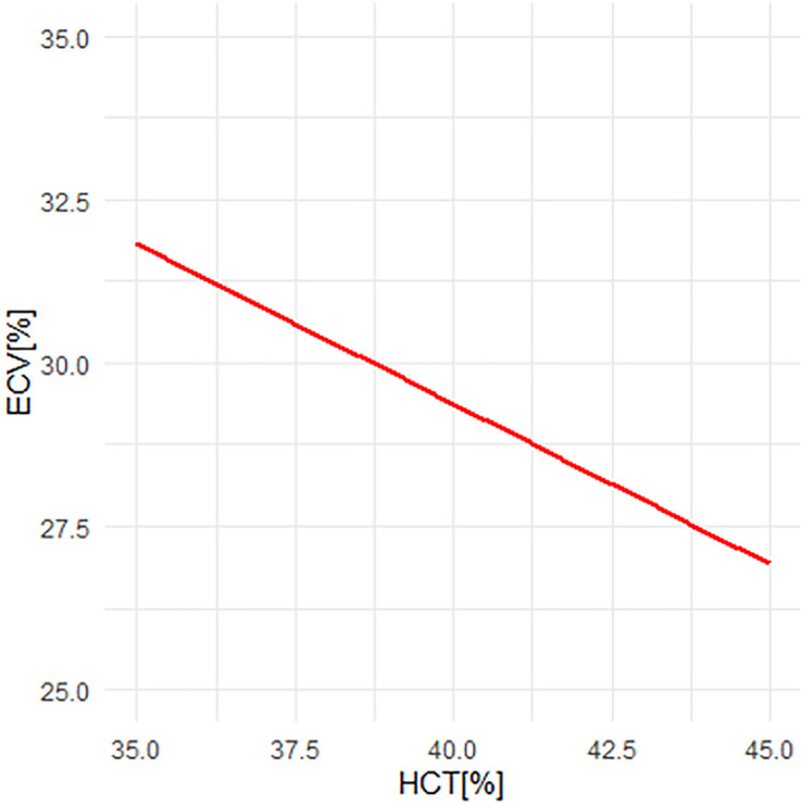
Cardiac magnetic resonance (CMR) 1 relaxation time mapping is an established technique primarily used to identify diffuse interstitial fibrosis and oedema. The myocardial extracellular volume (ECV) can be calculated from pre- and post-contrast 1 relaxation times and is a reproducible parametric index of the proportion of volume occupied by non-cardiomyocyte components in myocardial tissue. The conventional calculation of the ECV requires blood sampling to measure the haematocrit (HCT). Given the high variability of the HCT, the blood collection is recommended within 24 h of the CMR scan, limiting its applicability and posing a barrier to the clinical routine use of ECV measurements. In recent years, several research groups have proposed a method to determine the ECV by CMR without blood sampling. This is based on the inverse relationship between the 1 relaxation rate (1) of blood and the HCT. Consequently, a 'synthetic' HCT could be estimated from the native blood 1, avoiding blood sampling.

METHODS AND RESULTS

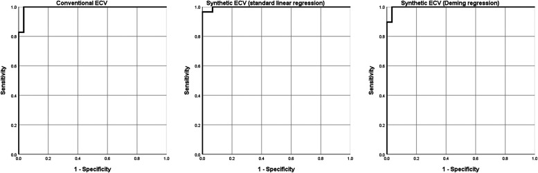
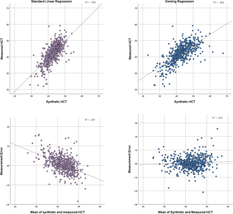
We performed a review and meta-analysis of published studies on synthetic ECV, as well as a secondary analysis of previously published data to examine the effect of the chosen regression modell on bias. While, overall, a good correlation and little bias between synthetic and conventional ECV were found in these studies, questions regarding its accuracy remain.

CONCLUSION

Synthetic HCT and ECV can provide a 'non-invasive' quantitative measurement of the myocardium's extracellular space when timely HCT measurements are not available and large alterations in ECV are expected, such as in cardiac amyloidosis. Due to the dependency of 1 relaxation times on the local setup, calculation of local formulas using linear regression is recommended, which can be easily performed using available data.

摘要

目的

心脏磁共振(CMR)T1弛豫时间映射是一种成熟的技术,主要用于识别弥漫性间质纤维化和水肿。心肌细胞外容积(ECV)可根据对比剂注射前后的T1弛豫时间计算得出,是心肌组织中非心肌细胞成分所占容积比例的一个可重复的参数指标。传统的ECV计算需要采集血样来测量血细胞比容(HCT)。鉴于HCT的高度变异性,建议在CMR扫描后24小时内采集血样,这限制了其适用性,并对ECV测量在临床常规应用中构成了障碍。近年来,几个研究小组提出了一种无需采集血样即可通过CMR测定ECV的方法。这是基于血液的T1弛豫率(R1)与HCT之间的反比关系。因此,可以根据天然血液的R1估算出“合成”HCT,从而避免采集血样。

方法和结果

我们对已发表的关于合成ECV的研究进行了综述和荟萃分析,并对先前发表的数据进行了二次分析,以检验所选回归模型对偏差的影响。虽然总体而言,这些研究发现合成ECV与传统ECV之间具有良好的相关性且偏差较小,但其准确性仍存在问题。

结论

当无法及时测量HCT且预计ECV会有较大变化时,如在心脏淀粉样变性中,合成HCT和ECV可以提供心肌细胞外间隙的“非侵入性”定量测量。由于T1弛豫时间依赖于局部设置,建议使用线性回归计算局部公式,这可以使用现有数据轻松完成。

https://cdn.ncbi.nlm.nih.gov/pmc/blobs/0b89/11195702/4d8ec4293d57/qyad022f3.jpg
https://cdn.ncbi.nlm.nih.gov/pmc/blobs/0b89/11195702/d8e556504057/qyad022_ga1.jpg
https://cdn.ncbi.nlm.nih.gov/pmc/blobs/0b89/11195702/461ca13caa0d/qyad022f1.jpg
https://cdn.ncbi.nlm.nih.gov/pmc/blobs/0b89/11195702/9fba271bf091/qyad022f2.jpg
https://cdn.ncbi.nlm.nih.gov/pmc/blobs/0b89/11195702/4d8ec4293d57/qyad022f3.jpg

相似文献

1
Quantification of myocardial extracellular volume without blood sampling.无需采血的心肌细胞外容积定量分析。
Eur Heart J Imaging Methods Pract. 2023 Sep 15;1(2):qyad022. doi: 10.1093/ehjimp/qyad022. eCollection 2023 Sep.
2
Establishment and validation of an extracellular volume model without blood sampling in ST-segment elevation myocardial infarction patients.ST段抬高型心肌梗死患者无需采血的细胞外容积模型的建立与验证
Eur Heart J Imaging Methods Pract. 2024 Jun 10;2(1):qyae053. doi: 10.1093/ehjimp/qyae053. eCollection 2024 Jan.
3
Automatic Measurement of the Myocardial Interstitium: Synthetic Extracellular Volume Quantification Without Hematocrit Sampling.心肌间质的自动测量:无需红细胞压积采样的合成细胞外体积定量。
JACC Cardiovasc Imaging. 2016 Jan;9(1):54-63. doi: 10.1016/j.jcmg.2015.11.008.
4
Myocardial extracellular volume fraction quantification in an animal model of the doxorubicin-induced myocardial fibrosis: a synthetic hematocrit method using 3T cardiac magnetic resonance.多柔比星诱导的心肌纤维化动物模型中心肌细胞外容积分数的定量分析:一种使用3T心脏磁共振成像的合成血细胞比容法
Quant Imaging Med Surg. 2021 Feb;11(2):510-520. doi: 10.21037/qims-20-501.
5
Extracellular volume quantification by cardiac magnetic resonance imaging without hematocrit sampling : Ready for prime time?无需采集血细胞比容的心脏磁共振成像细胞外容积定量:已准备好进入黄金时代了吗?
Wien Klin Wochenschr. 2018 Mar;130(5-6):190-196. doi: 10.1007/s00508-017-1267-y. Epub 2017 Oct 4.
6
Synthetic hematocrit derived from the longitudinal relaxation of blood can lead to clinically significant errors in measurement of extracellular volume fraction in pediatric and young adult patients.源自血液纵向弛豫的合成血细胞比容可能导致儿科和年轻成年患者细胞外体积分数测量出现具有临床意义的误差。
J Cardiovasc Magn Reson. 2017 Aug 2;19(1):58. doi: 10.1186/s12968-017-0377-z.
7
Estimating synthetic hematocrit and extracellular volume from native blood pool T1 times at 3 Tesla CMR: Derivation of a conversion equation, accuracy and comparison with published formulas.从 3T CMR 中的原生血池 T1 时间估算合成血细胞比容和细胞外容积:转换方程的推导、准确性及与已发表公式的比较。
Eur J Radiol. 2024 Sep;178:111659. doi: 10.1016/j.ejrad.2024.111659. Epub 2024 Jul 31.
8
Automatic quantification of the myocardial extracellular volume by cardiac computed tomography: Synthetic ECV by CCT.通过心脏计算机断层扫描自动定量心肌细胞外容积:心脏CT合成细胞外容积(ECV)
J Cardiovasc Comput Tomogr. 2017 May-Jun;11(3):221-226. doi: 10.1016/j.jcct.2017.02.006. Epub 2017 Feb 22.
9
Synthetic extracellular volume fraction without hematocrit sampling for hepatic applications.无红细胞压积采样的合成细胞外体积分数在肝脏应用中的应用。
Abdom Radiol (NY). 2021 Oct;46(10):4637-4646. doi: 10.1007/s00261-021-03140-6. Epub 2021 Jun 10.
10
Is a timely assessment of the hematocrit necessary for cardiovascular magnetic resonance-derived extracellular volume measurements?是否需要及时评估血细胞比容以进行心血管磁共振衍生的细胞外容积测量?
J Cardiovasc Magn Reson. 2020 Nov 30;22(1):77. doi: 10.1186/s12968-020-00689-x.

引用本文的文献

1
Establishment of a synthetic ECV model and its prognostic value in diabetes patients with acute myocardial infarction.建立合成的内皮祖细胞模型及其在糖尿病急性心肌梗死患者中的预后价值。
Front Endocrinol (Lausanne). 2025 Jun 25;16:1534236. doi: 10.3389/fendo.2025.1534236. eCollection 2025.

本文引用的文献

1
Synthetic Extracellular Volume in Cardiac Magnetic Resonance Without Blood Sampling: a Reliable Tool to Replace Conventional Extracellular Volume.心脏磁共振无采血的合成细胞外容积:替代常规细胞外容积的可靠工具。
Circ Cardiovasc Imaging. 2022 Apr;15(4):e013745. doi: 10.1161/CIRCIMAGING.121.013745. Epub 2022 Apr 1.
2
Amyloid Deposits and Fibrosis on Left Ventricular Endomyocardial Biopsy Correlate With Extracellular Volume in Cardiac Amyloidosis.左心室心肌内膜活检中的淀粉样沉积物和纤维化与心脏淀粉样变性中的细胞外容积相关。
J Am Heart Assoc. 2021 Oct 19;10(20):e020358. doi: 10.1161/JAHA.120.020358. Epub 2021 Oct 8.
3
T1 Mapping and Extracellular Volume Fraction in Dilated Cardiomyopathy: A Prognosis Study.
T1 映射和扩张型心肌病中的细胞外容积分数:一项预后研究。
JACC Cardiovasc Imaging. 2022 Apr;15(4):578-590. doi: 10.1016/j.jcmg.2021.07.023. Epub 2021 Sep 15.
4
Reference ranges ("normal values") for cardiovascular magnetic resonance (CMR) in adults and children: 2020 update.成人和儿童心血管磁共振(CMR)的参考范围(“正常值”):2020 更新。
J Cardiovasc Magn Reson. 2020 Dec 14;22(1):87. doi: 10.1186/s12968-020-00683-3.
5
Is a timely assessment of the hematocrit necessary for cardiovascular magnetic resonance-derived extracellular volume measurements?是否需要及时评估血细胞比容以进行心血管磁共振衍生的细胞外容积测量?
J Cardiovasc Magn Reson. 2020 Nov 30;22(1):77. doi: 10.1186/s12968-020-00689-x.
6
Minimum sample size for external validation of a clinical prediction model with a continuous outcome.用于连续结局临床预测模型外部验证的最小样本量。
Stat Med. 2021 Jan 15;40(1):133-146. doi: 10.1002/sim.8766. Epub 2020 Nov 4.
7
Myofibroblasts and Fibrosis: Mitochondrial and Metabolic Control of Cellular Differentiation.肌成纤维细胞与纤维化:细胞分化的线粒体与代谢调控
Circ Res. 2020 Jul 17;127(3):427-447. doi: 10.1161/CIRCRESAHA.120.316958. Epub 2020 Jul 16.
8
Calculating the sample size required for developing a clinical prediction model.计算开发临床预测模型所需的样本量。
BMJ. 2020 Mar 18;368:m441. doi: 10.1136/bmj.m441.
9
Left ventricular fibrosis by extracellular volume fraction and the risk of atrial fibrillation recurrence after catheter ablation.基于细胞外容积分数的左心室纤维化与导管消融术后房颤复发风险
Cardiovasc Diagn Ther. 2019 Dec;9(6):578-585. doi: 10.21037/cdt.2019.12.03.
10
Myocardial T1 and ECV Measurement: Underlying Concepts and Technical Considerations.心肌 T1 和 ECV 测量:基本概念和技术考虑因素。
JACC Cardiovasc Imaging. 2019 Nov;12(11 Pt 2):2332-2344. doi: 10.1016/j.jcmg.2019.06.031. Epub 2019 Sep 18.