Department of Biomedical Engineering, Vanderbilt University, Nashville, Tennessee, USA.
Vanderbilt University Institute of Imaging Science, Vanderbilt University Medical Center, Nashville, Tennessee, USA.
Epilepsia. 2024 Sep;65(9):2686-2699. doi: 10.1111/epi.18074. Epub 2024 Jul 26.
Epilepsy is a common neurological disorder affecting 1% of the global population. Loss of consciousness in focal impaired awareness seizures (FIASs) and focal-to-bilateral tonic-clonic seizures (FBTCSs) can be devastating, but the mechanisms are not well understood. Although ictal activity and interictal connectivity changes have been noted, the network states of focal aware seizures (FASs), FIASs, and FBTCSs have not been thoroughly evaluated with network measures ictally.
We obtained electrographic data from 74 patients with stereoelectroencephalography (SEEG). Sliding window band power, functional connectivity, and segregation were computed on preictal, ictal, and postictal data. Five-minute epochs of wake, rapid eye movement sleep, and deep sleep were also extracted. Connectivity of subcortical arousal structures was analyzed in a cohort of patients with both SEEG and functional magnetic resonance imaging (fMRI). Given that custom neuromodulation of seizures is predicated on detection of seizure type, a convolutional neural network was used to classify seizure types.
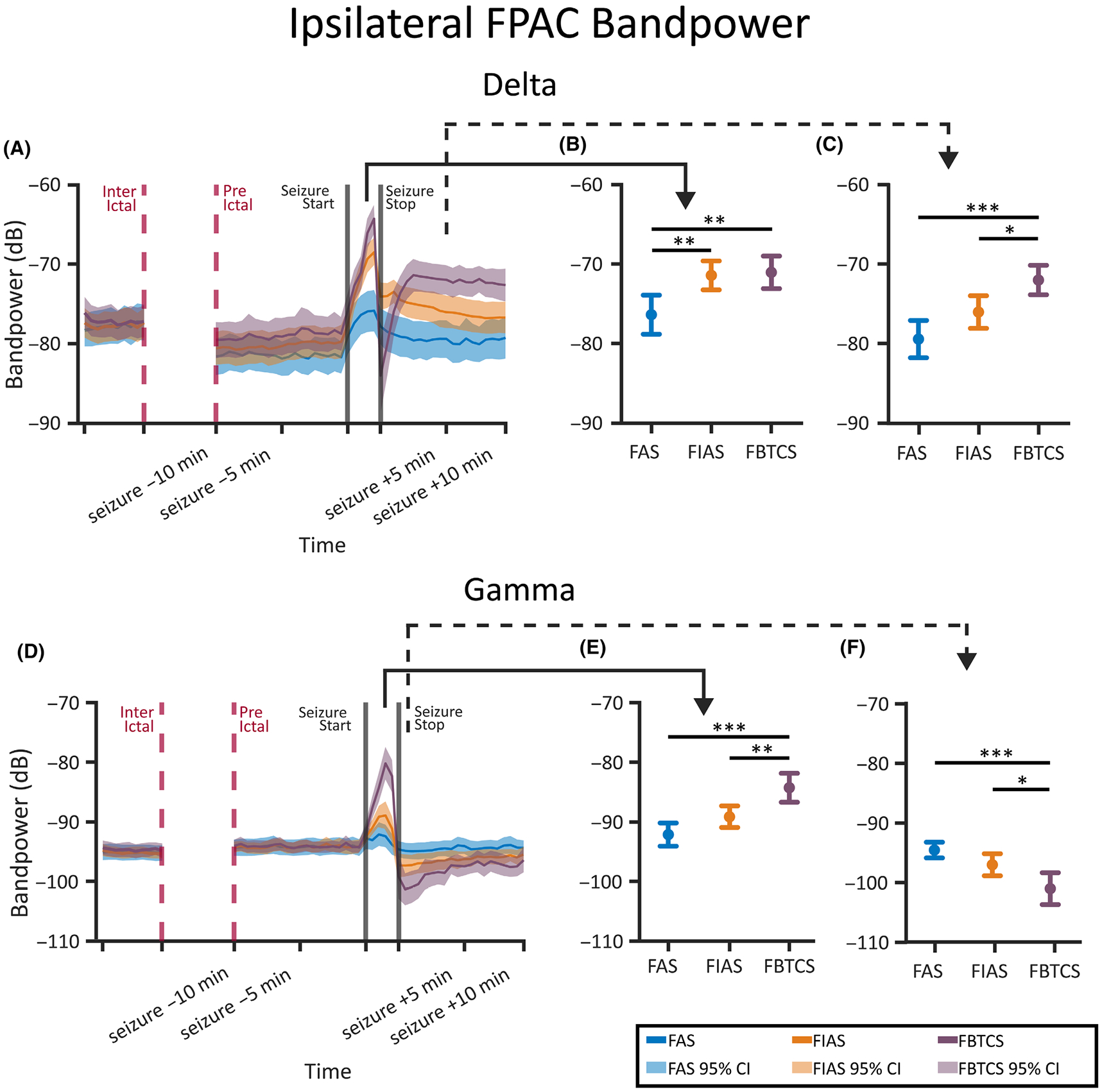
We found that in the frontoparietal association cortex, an area associated with consciousness, both consciousness-impairing seizures (FIASs and FBTCSs) and deep sleep had increases in slow wave delta (1-4 Hz) band power. However, when network measures were employed, we found that only FIASs and deep sleep exhibited an increase in delta segregation and a decrease in gamma segregation. Furthermore, we found that only patients with FIASs had reduced subcortical-to-neocortical functional connectivity with fMRI versus controls. Finally, our deep learning network demonstrated an area under the curve of .75 for detecting consciousness-impairing seizures.
This study provides novel insights into ictal network measures in FASs, FIASs, and FBTCSs. Importantly, although both FIASs and FBTCSs result in loss of consciousness, our results suggest that ictal network changes in FIASs uniquely resemble those that occur during deep sleep. Our results may inform novel neuromodulation strategies for preservation of consciousness in epilepsy.
癫痫是一种常见的神经系统疾病,影响全球 1%的人口。局灶性意识障碍发作(FIASs)和局灶性双侧强直阵挛发作(FBTCSs)引起的意识丧失可能是毁灭性的,但机制尚不清楚。尽管已经注意到发作期活动和发作间期连接变化,但尚未使用网络测量方法全面评估局灶性意识发作(FASs)、FIASs 和 FBTCSs 的网络状态。
我们从 74 例立体脑电图(SEEG)患者中获得了脑电图数据。在发作前、发作期和发作后数据上计算了滑动窗口频带功率、功能连接和分离。还提取了清醒、快速眼动睡眠和深度睡眠的 5 分钟时程。分析了具有 SEEG 和功能磁共振成像(fMRI)的患者亚皮质唤醒结构的连接。鉴于对癫痫发作的定制神经调节取决于对癫痫发作类型的检测,因此使用卷积神经网络来对癫痫发作类型进行分类。
我们发现,在前顶联合皮层,一个与意识相关的区域,意识障碍性发作(FIASs 和 FBTCSs)和深度睡眠都增加了慢波 delta(1-4 Hz)频带功率。然而,当使用网络测量方法时,我们发现只有 FIASs 和深度睡眠表现出 delta 分离增加和 gamma 分离减少。此外,我们发现只有 FIASs 患者与对照组相比,fMRI 显示亚皮质到新皮质的功能连接减少。最后,我们的深度学习网络对意识障碍性癫痫发作的检测曲线下面积为 0.75。
本研究提供了 FASs、FIASs 和 FBTCSs 发作期网络测量的新见解。重要的是,尽管 FIASs 和 FBTCSs 都导致意识丧失,但我们的结果表明,FIASs 中的发作期网络变化与深度睡眠期间发生的变化具有独特的相似性。我们的研究结果可能为癫痫患者意识保护的新型神经调节策略提供信息。