Millen Aletta M E, Maluleke Tshiamo T, Pienaar Leandrie, Sallie Farhanah N, Veerappan Radhini, Andrén Per E, Baijnath Sooraj
Wits Integrated Molecular Physiology Research Initiative, Wits Health Consortium (PTY) Ltd., University of the Witwatersrand, Johannesburg 2191, South Africa.
School of Physiology, Faculty of Health Sciences, University of The Witwatersrand, Johannesburg 2191, South Africa.
Biology (Basel). 2024 Jul 10;13(7):516. doi: 10.3390/biology13070516.
The effects of collagen-induced arthritis (CIA), a model of systemic inflammation, on brain regional molecular markers associated with neurological disorders are uncertain.
This study investigated the brain regional molecular changes in markers associated with inflammation and neuronal dysfunction in a CIA model.
Fourteen male Sprague Dawley rats were divided into control (n = 5) or CIA (n = 9) groups. 10 weeks after CIA induction, brain tissue was collected. Brain regional mRNA expression of inflammatory markers ( and ), apoptotic markers ( and ) and neurotrophic factors (, and ) was determined. Monoamine distribution and abundance in different brain regions were determine by mass spectrometry imaging (MSI).
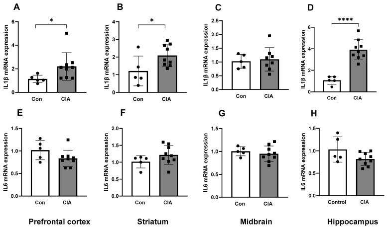
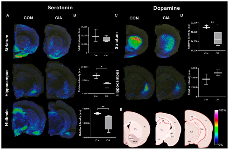
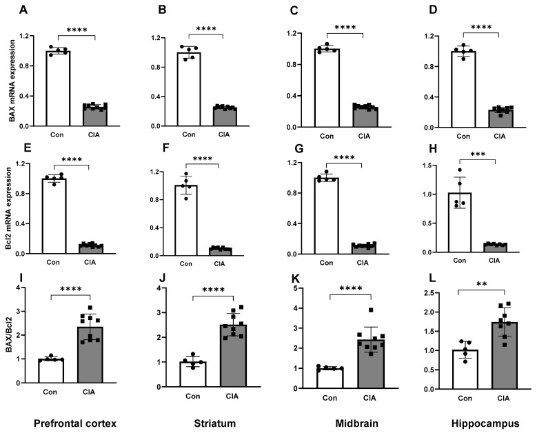
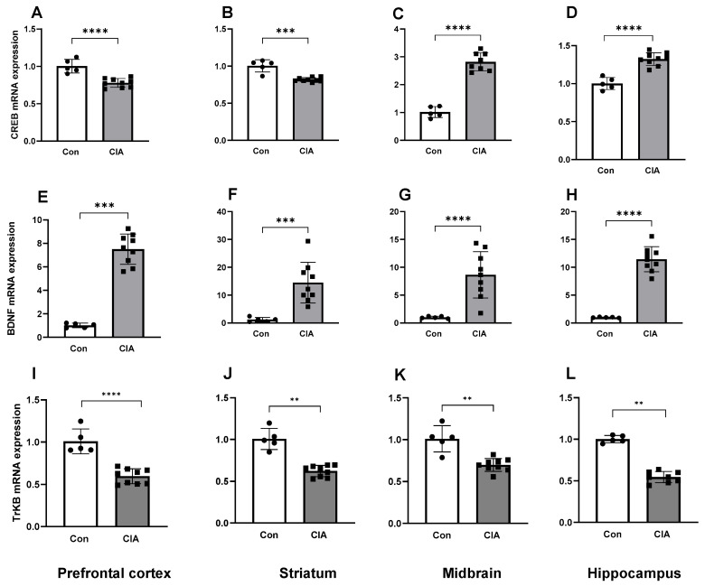
Neuroinflammation was confirmed in the CIA group by increased mRNA expression, concurrent with an increased ratio. The mRNA expression of was increased in the midbrain and hippocampus while was increased and was decreased across all brain regions in CIA compared to control animals. Serotonin was decreased in the midbrain and hippocampus while dopamine was decreased in the striatum of CIA rats, compared to controls.
CIA resulted in neuroinflammation concurrent with an apoptotic state and aberrant expression of neurotrophic factors and monoamines in the brain, suggestive of neurodegeneration.
胶原诱导的关节炎(CIA)作为一种全身炎症模型,其对与神经疾病相关的脑区分子标志物的影响尚不确定。
本研究在CIA模型中探究与炎症和神经元功能障碍相关的标志物在脑区的分子变化。
将14只雄性Sprague Dawley大鼠分为对照组(n = 5)和CIA组(n = 9)。诱导CIA 10周后,收集脑组织。测定脑区炎症标志物(和)、凋亡标志物(和)以及神经营养因子(、和)的mRNA表达。通过质谱成像(MSI)确定不同脑区单胺的分布和丰度。
CIA组中,mRNA表达增加证实了神经炎症,同时比值升高。与对照动物相比,CIA组中脑和海马体中的mRNA表达增加,而所有脑区中的增加,减少。与对照组相比,CIA大鼠中脑和海马体中的5-羟色胺减少,纹状体中的多巴胺减少。
CIA导致神经炎症,同时伴有凋亡状态以及脑内神经营养因子和单胺的异常表达,提示神经退行性变。