Calandrelli Rosalinda, D'Apolito Gabriella, Martucci Matia, Giordano Carolina, Schiarelli Chiara, Marziali Giammaria, Varcasia Giuseppe, Ausili Cefaro Luca, Chiloiro Sabrina, De Sanctis Simone Antonio, Serioli Simona, Doglietto Francesco, Gaudino Simona
Department of Imaging, Radiation Therapy and Hematology, Catholic University of the Sacred Heart, Fondazione Policlinico Universitario A. Gemelli IRCCS, 00168 Rome, Italy.
Pituitary Unit, Division of Endocrinology and Metabolism, Fondazione Policlinico Universitario A. Gemelli, IRCCS, 00168 Rome, Italy.
Cancers (Basel). 2024 Jul 13;16(14):2532. doi: 10.3390/cancers16142532.
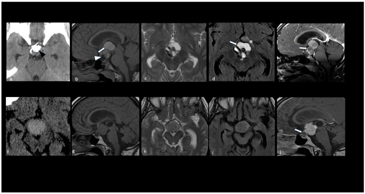
Craniopharyngiomas continue to present a challenge in clinical practice due to their heterogeneity and unpredictable adherence to vital neurovascular structures, particularly the hypothalamus. This results in different degrees of hypothalamus-pituitary axis dysfunction and a lack of uniform consensus and treatment guidelines regarding optimal management. MRI and CT are complementary techniques in the preoperative diagnostic phase, enabling the precise definition of craniopharyngioma size, shape, and consistency, as well as guiding classification into histopathological subtypes and topographical categories. Meanwhile, MRI plays a crucial role in the immediate postoperative period and follow-up stages by identifying treatment-related changes and residual tumors. This pictorial essay aims to provide an overview of the role of imaging in identifying variables indicative of the adherence degree to the hypothalamus, hypothalamic-pituitary dysfunction, the extent of surgical excision, and prognosis. For a more comprehensive assessment, we choose to distinguish the following two scenarios: (1) the initial diagnosis phase, where we primarily discuss the role of radiological variables predictive of adhesions to the surrounding neurovascular structures and axis dysfunction which may influence the choice of surgical resection; (2) the early post-treatment follow-up phase, where we discuss the interpretation of treatment-related changes that impact outcomes.
颅咽管瘤因其异质性以及对重要神经血管结构(尤其是下丘脑)的不可预测的粘连性,在临床实践中仍然是一个挑战。这导致不同程度的下丘脑 - 垂体轴功能障碍,并且在最佳治疗管理方面缺乏统一的共识和治疗指南。在术前诊断阶段,磁共振成像(MRI)和计算机断层扫描(CT)是互补技术,能够精确界定颅咽管瘤的大小、形状和质地,还能指导其组织病理学亚型和地形学分类。同时,MRI在术后即刻及随访阶段起着关键作用,可识别与治疗相关的变化及残留肿瘤。这篇图文并茂的文章旨在概述影像学在识别提示与下丘脑粘连程度、下丘脑 - 垂体功能障碍、手术切除范围及预后的变量方面的作用。为了进行更全面的评估,我们选择区分以下两种情况:(1)初始诊断阶段,在此我们主要讨论预测与周围神经血管结构粘连及可能影响手术切除选择的轴功能障碍的放射学变量的作用;(2)治疗后早期随访阶段,在此我们讨论影响治疗结果的与治疗相关变化的解读。