• 文献检索
  • 文档翻译
  • 深度研究
  • 学术资讯
  • Suppr Zotero 插件Zotero 插件
  • 邀请有礼
  • 套餐&价格
  • 历史记录
应用&插件
Suppr Zotero 插件Zotero 插件浏览器插件Mac 客户端Windows 客户端微信小程序
定价
高级版会员购买积分包购买API积分包
服务
文献检索文档翻译深度研究API 文档MCP 服务
关于我们
关于 Suppr公司介绍联系我们用户协议隐私条款
关注我们

Suppr 超能文献

核心技术专利:CN118964589B侵权必究
粤ICP备2023148730 号-1Suppr @ 2026

文献检索

告别复杂PubMed语法,用中文像聊天一样搜索,搜遍4000万医学文献。AI智能推荐,让科研检索更轻松。

立即免费搜索

文件翻译

保留排版,准确专业,支持PDF/Word/PPT等文件格式,支持 12+语言互译。

免费翻译文档

深度研究

AI帮你快速写综述,25分钟生成高质量综述,智能提取关键信息,辅助科研写作。

立即免费体验

妊娠11至14周二维胎盘生物测量和三维胎盘体积的参考范围。

Reference Ranges of 2-Dimensional Placental Biometry and 3-Dimensional Placental Volume between 11 and 14 Weeks of Gestation.

作者信息

Trilla Solà Cristina, Parra Roca Juan, Llurba Olivé Elisa

机构信息

Department of Obstetrics and Gynecology, Institut d'Investigació Biomèdica Sant Pau-IIB Sant Pau, Hospital de la Santa Creu i Sant Pau, 08025 Barcelona, Spain.

Faculty of Medicine, Universitat Autònoma de Barcelona, 08025 Barcelona, Spain.

出版信息

Diagnostics (Basel). 2024 Jul 18;14(14):1556. doi: 10.3390/diagnostics14141556.

DOI:10.3390/diagnostics14141556
PMID:39061694
原文链接:https://pmc.ncbi.nlm.nih.gov/articles/PMC11276113/
Abstract

OBJECTIVE

The purpose of this study was to provide gestational age (GA) specific reference ranges for 2-dimensional (2D) placental biometry and 3-dimensional (3D) placental volume between 11 and 14 weeks of gestation.

METHODS

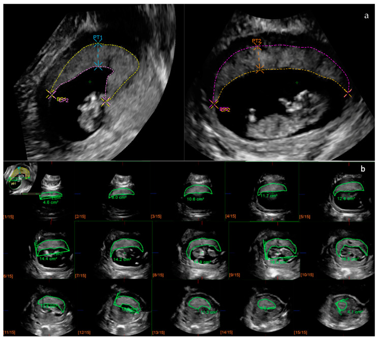
Placental biometry including 2D and 3D variables was calculated in 1142 first-trimester singleton pregnancies with non-complicated outcome between September 2016 and February 2020. Ultrasound datasets were obtained at the time of the first-trimester ultrasound, and 2D basal plate (BP), chorionic plate (CP), placental thickness (PT), and 3D placental volume (PV) were measured following a standardized methodology. Reference ranges for each variable were calculated according to GA and crown-rump-length (CRL).

RESULTS

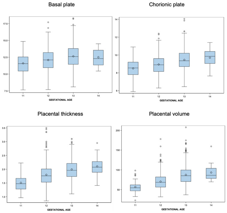
A total of 1142 uncomplicated pregnancies were considered for analysis. All placental measurements increased significantly between 11 and 14 weeks, especially for PT (39.64%) and PV (64.4%). Reference ranges were constructed for each 2D and 3D first-trimester placental variable using the best-fit regression model for the predicted mean and SD as a function of GA and CRL.

CONCLUSIONS

Reference ranges of 2D placental biometry and 3D placental volume between 11 and 14 weeks of gestation were constructed, generating reference values. Placental biometry showed a progressive increase during the first trimester. This highlights the importance of using reference range charts according to GA.

摘要

目的

本研究旨在提供妊娠11至14周二维(2D)胎盘生物测量和三维(3D)胎盘体积的特定孕周(GA)参考范围。

方法

对2016年9月至2020年2月期间1142例孕早期单胎妊娠且结局无并发症的孕妇进行胎盘生物测量,包括2D和3D变量。在孕早期超声检查时获取超声数据集,并按照标准化方法测量2D基底板(BP)、绒毛板(CP)、胎盘厚度(PT)和3D胎盘体积(PV)。根据孕周和头臀长(CRL)计算每个变量的参考范围。

结果

共1142例无并发症妊娠纳入分析。所有胎盘测量值在11至14周之间均显著增加,尤其是PT(39.64%)和PV(64.4%)。使用最佳拟合回归模型,以孕周和CRL的函数形式预测均值和标准差,为孕早期每个2D和3D胎盘变量构建参考范围。

结论

构建了妊娠11至14周2D胎盘生物测量和3D胎盘体积的参考范围,得出了参考值。胎盘生物测量在孕早期呈逐渐增加趋势。这突出了根据孕周使用参考范围图表的重要性。

https://cdn.ncbi.nlm.nih.gov/pmc/blobs/c46f/11276113/f406118c050c/diagnostics-14-01556-g002.jpg
https://cdn.ncbi.nlm.nih.gov/pmc/blobs/c46f/11276113/c497794ff40d/diagnostics-14-01556-g001.jpg
https://cdn.ncbi.nlm.nih.gov/pmc/blobs/c46f/11276113/f406118c050c/diagnostics-14-01556-g002.jpg
https://cdn.ncbi.nlm.nih.gov/pmc/blobs/c46f/11276113/c497794ff40d/diagnostics-14-01556-g001.jpg
https://cdn.ncbi.nlm.nih.gov/pmc/blobs/c46f/11276113/f406118c050c/diagnostics-14-01556-g002.jpg

相似文献

1
Reference Ranges of 2-Dimensional Placental Biometry and 3-Dimensional Placental Volume between 11 and 14 Weeks of Gestation.妊娠11至14周二维胎盘生物测量和三维胎盘体积的参考范围。
Diagnostics (Basel). 2024 Jul 18;14(14):1556. doi: 10.3390/diagnostics14141556.
2
Inter- and Intraobserver Agreement in First Trimester Ultrasound Evaluation of Placental Biometry.孕早期胎盘生物测量超声评估中的观察者间和观察者内一致性
Fetal Diagn Ther. 2020;47(11):834-840. doi: 10.1159/000509707. Epub 2020 Aug 12.
3
Reference ranges of placental volume measured by virtual organ computer-aided analysis between 10 and 14 weeks of gestation.孕10至14周期间通过虚拟器官计算机辅助分析测量的胎盘体积参考范围。
J Clin Ultrasound. 2017 May;45(4):185-191. doi: 10.1002/jcu.22441. Epub 2017 Feb 6.
4
Third trimester placental volume and biometry measurement: A method-development study.孕晚期胎盘体积和生物测量:一项方法开发研究。
Placenta. 2016 Jun;42:51-8. doi: 10.1016/j.placenta.2016.04.010. Epub 2016 Apr 9.
5
First trimester brain ventricle fluid and embryonic volumes measured by three-dimensional ultrasound with the use of I-Space virtual reality.应用 I-Space 虚拟现实技术的三维超声测量头三个月脑室液和胚胎体积。
Hum Reprod. 2013 May;28(5):1181-9. doi: 10.1093/humrep/det035. Epub 2013 Mar 6.
6
Reference ranges and distribution of placental volume by 3-dimensional virtual organ computer-aided analysis between 11 weeks and 13 weeks 6 days.11 周至 13 周 6 天期间通过三维虚拟器官计算机辅助分析的胎盘容量参考范围和分布。
J Ultrasound Med. 2013 Aug;32(8):1477-82. doi: 10.7863/ultra.32.8.1477.
7
International standards for early fetal size and pregnancy dating based on ultrasound measurement of crown-rump length in the first trimester of pregnancy.基于孕早期超声测量头臀长的早期胎儿大小和孕周确定的国际标准。
Ultrasound Obstet Gynecol. 2014 Dec;44(6):641-8. doi: 10.1002/uog.13448. Epub 2014 Nov 2.
8
First-trimester utero-placental (vascular) development and embryonic and fetal growth: The Rotterdam periconception cohort.早孕期子宫胎盘(血管)发育及胚胎和胎儿生长:鹿特丹围孕期队列研究。
Placenta. 2021 May;108:81-90. doi: 10.1016/j.placenta.2021.03.017. Epub 2021 Mar 28.
9
Three-dimensional placental volume and mean grey value: Normal ranges in a Turkish population and correlation with maternal serum biochemistry and Doppler parameters.三维胎盘体积和平均灰度值:土耳其人群的正常范围及其与母体血清生化指标和多普勒参数的相关性
J Obstet Gynaecol. 2015 Apr;35(3):259-62. doi: 10.3109/01443615.2014.958146. Epub 2014 Sep 25.
10
Placental volume measured by three-dimensional ultrasound in the prediction of fetal alpha(0)-thalassemia: a preliminary report.三维超声测量胎盘体积预测胎儿α(0)地中海贫血:初步报告
Ultrasound Obstet Gynecol. 2006 Aug;28(2):166-72. doi: 10.1002/uog.2721.

引用本文的文献

1
Reference charts for first-trimester placental volume derived using OxNNet.使用OxNNet得出的孕早期胎盘体积参考图表。
Ultrasound Obstet Gynecol. 2025 Sep;66(3):337-346. doi: 10.1002/uog.29300. Epub 2025 Aug 1.

本文引用的文献

1
The PURPOSe cause of death study in stillbirths and neonatal deaths in India and Pakistan: A review.印度和巴基斯坦死产和新生儿死亡的 PURPOSE 死因研究:综述。
BJOG. 2023 Nov;130 Suppl 3:26-35. doi: 10.1111/1471-0528.17635. Epub 2023 Aug 17.
2
Pregnancy Complications, Correlation With Placental Pathology and Neonatal Outcomes.妊娠并发症、与胎盘病理学及新生儿结局的相关性
Front Clin Diabetes Healthc. 2022 Mar 8;2:807192. doi: 10.3389/fcdhc.2021.807192. eCollection 2021.
3
New Frontiers in Placenta Tissue Imaging.胎盘组织成像的新前沿
EMJ Radiol. 2020 Sep;1(1):54-62. doi: 10.33590/emjradiol/19-00210.
4
Human placental development and function.人类胎盘的发育和功能。
Semin Cell Dev Biol. 2022 Nov;131:66-77. doi: 10.1016/j.semcdb.2022.03.039. Epub 2022 Apr 4.
5
Inter- and Intraobserver Agreement in First Trimester Ultrasound Evaluation of Placental Biometry.孕早期胎盘生物测量超声评估中的观察者间和观察者内一致性
Fetal Diagn Ther. 2020;47(11):834-840. doi: 10.1159/000509707. Epub 2020 Aug 12.
6
Development of the human placenta.人类胎盘的发育。
Development. 2019 Nov 27;146(22):dev163428. doi: 10.1242/dev.163428.
7
Inter-observer agreement and reliability assessment for observational studies of clinical work.临床工作观察性研究的观察者间一致性和可靠性评估。
J Biomed Inform. 2019 Dec;100:103317. doi: 10.1016/j.jbi.2019.103317. Epub 2019 Oct 22.
8
Early development of the human placenta and pregnancy complications.人类胎盘的早期发育与妊娠并发症。
J Matern Fetal Neonatal Med. 2020 Oct;33(20):3538-3545. doi: 10.1080/14767058.2019.1578745. Epub 2019 Feb 27.
9
First trimester 3D ultrasound placental volume for predicting preeclampsia and/or intrauterine growth restriction.孕早期三维超声胎盘体积预测子痫前期和/或胎儿生长受限。
J Obstet Gynaecol. 2019 May;39(4):474-479. doi: 10.1080/01443615.2018.1529152. Epub 2018 Dec 25.
10
Pathophysiology of placental-derived fetal growth restriction.胎盘源性胎儿生长受限的病理生理学。
Am J Obstet Gynecol. 2018 Feb;218(2S):S745-S761. doi: 10.1016/j.ajog.2017.11.577.