• 文献检索
  • 文档翻译
  • 深度研究
  • 学术资讯
  • Suppr Zotero 插件Zotero 插件
  • 邀请有礼
  • 套餐&价格
  • 历史记录
应用&插件
Suppr Zotero 插件Zotero 插件浏览器插件Mac 客户端Windows 客户端微信小程序
定价
高级版会员购买积分包购买API积分包
服务
文献检索文档翻译深度研究API 文档MCP 服务
关于我们
关于 Suppr公司介绍联系我们用户协议隐私条款
关注我们

Suppr 超能文献

核心技术专利:CN118964589B侵权必究
粤ICP备2023148730 号-1Suppr @ 2026

文献检索

告别复杂PubMed语法,用中文像聊天一样搜索,搜遍4000万医学文献。AI智能推荐,让科研检索更轻松。

立即免费搜索

文件翻译

保留排版,准确专业,支持PDF/Word/PPT等文件格式,支持 12+语言互译。

免费翻译文档

深度研究

AI帮你快速写综述,25分钟生成高质量综述,智能提取关键信息,辅助科研写作。

立即免费体验

单细胞分辨率下小鼠大脑的时空转录组分析与建模揭示衰老和年轻化的细胞邻近效应

Spatiotemporal transcriptomic profiling and modeling of mouse brain at single-cell resolution reveals cell proximity effects of aging and rejuvenation.

作者信息

Sun Eric D, Zhou Olivia Y, Hauptschein Max, Rappoport Nimrod, Xu Lucy, Navarro Negredo Paloma, Liu Ling, Rando Thomas A, Zou James, Brunet Anne

机构信息

Department of Biomedical Data Science, Stanford University, CA, USA.

Department of Genetics, Stanford University, CA, USA.

出版信息

bioRxiv. 2024 Jul 19:2024.07.16.603809. doi: 10.1101/2024.07.16.603809.

DOI:10.1101/2024.07.16.603809
PMID:39071282
原文链接:https://pmc.ncbi.nlm.nih.gov/articles/PMC11275735/
Abstract

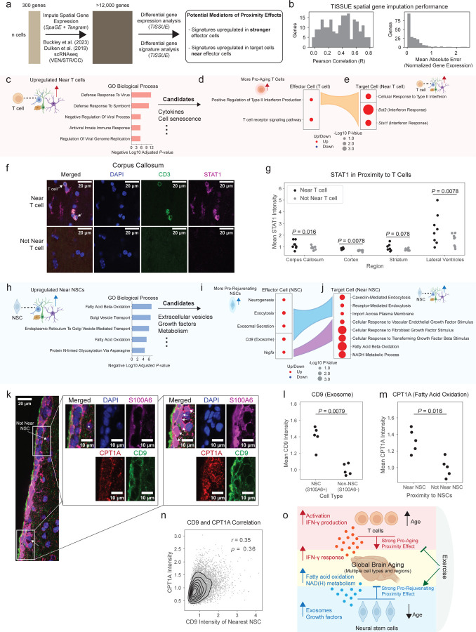
Old age is associated with a decline in cognitive function and an increase in neurodegenerative disease risk. Brain aging is complex and accompanied by many cellular changes. However, the influence that aged cells have on neighboring cells and how this contributes to tissue decline is unknown. More generally, the tools to systematically address this question in aging tissues have not yet been developed. Here, we generate spatiotemporal data at single-cell resolution for the mouse brain across lifespan, and we develop the first machine learning models based on spatial transcriptomics ('spatial aging clocks') to reveal cell proximity effects during brain aging and rejuvenation. We collect a single-cell spatial transcriptomics brain atlas of 4.2 million cells from 20 distinct ages and across two rejuvenating interventions-exercise and partial reprogramming. We identify spatial and cell type-specific transcriptomic fingerprints of aging, rejuvenation, and disease, including for rare cell types. Using spatial aging clocks and deep learning models, we find that T cells, which infiltrate the brain with age, have a striking pro-aging proximity effect on neighboring cells. Surprisingly, neural stem cells have a strong pro-rejuvenating effect on neighboring cells. By developing computational tools to identify mediators of these proximity effects, we find that pro-aging T cells trigger a local inflammatory response likely via interferon-γ whereas pro-rejuvenating neural stem cells impact the metabolism of neighboring cells possibly via growth factors (e.g. vascular endothelial growth factor) and extracellular vesicles, and we experimentally validate some of these predictions. These results suggest that rare cells can have a drastic influence on their neighbors and could be targeted to counter tissue aging. We anticipate that these spatial aging clocks will not only allow scalable assessment of the efficacy of interventions for aging and disease but also represent a new tool for studying cell-cell interactions in many spatial contexts.

摘要

衰老与认知功能衰退以及神经退行性疾病风险增加相关。大脑衰老过程复杂,伴随着许多细胞变化。然而,衰老细胞对邻近细胞的影响以及这种影响如何导致组织衰退尚不清楚。更普遍地说,在衰老组织中系统解决这个问题的工具尚未开发出来。在这里,我们生成了小鼠大脑在整个生命周期内单细胞分辨率的时空数据,并开发了首个基于空间转录组学的机器学习模型(“空间衰老时钟”),以揭示大脑衰老和恢复活力过程中的细胞邻近效应。我们收集了来自20个不同年龄以及两种恢复活力干预措施(运动和部分重编程)的420万个细胞的单细胞空间转录组学大脑图谱。我们确定了衰老、恢复活力和疾病的空间及细胞类型特异性转录组指纹,包括罕见细胞类型。使用空间衰老时钟和深度学习模型,我们发现随着年龄增长渗入大脑的T细胞对邻近细胞具有显著的促衰老邻近效应。令人惊讶的是,神经干细胞对邻近细胞具有强大的促恢复活力作用。通过开发计算工具来识别这些邻近效应的介导因子,我们发现促衰老T细胞可能通过干扰素-γ触发局部炎症反应,而促恢复活力的神经干细胞可能通过生长因子(如血管内皮生长因子)和细胞外囊泡影响邻近细胞的代谢,并且我们通过实验验证了其中一些预测。这些结果表明,罕见细胞可以对其邻居产生巨大影响,并且可以成为对抗组织衰老的靶点。我们预计这些空间衰老时钟不仅将允许对衰老和疾病干预措施的疗效进行可扩展评估,而且还将成为研究许多空间背景下细胞间相互作用的新工具。

https://cdn.ncbi.nlm.nih.gov/pmc/blobs/32dd/11275735/dd5e55a21ed3/nihpp-2024.07.16.603809v1-f0015.jpg
https://cdn.ncbi.nlm.nih.gov/pmc/blobs/32dd/11275735/0085f232043e/nihpp-2024.07.16.603809v1-f0001.jpg
https://cdn.ncbi.nlm.nih.gov/pmc/blobs/32dd/11275735/9fccc1a8c25e/nihpp-2024.07.16.603809v1-f0002.jpg
https://cdn.ncbi.nlm.nih.gov/pmc/blobs/32dd/11275735/a5600763167d/nihpp-2024.07.16.603809v1-f0003.jpg
https://cdn.ncbi.nlm.nih.gov/pmc/blobs/32dd/11275735/bab7779e7e4b/nihpp-2024.07.16.603809v1-f0004.jpg
https://cdn.ncbi.nlm.nih.gov/pmc/blobs/32dd/11275735/44a9d6f221c7/nihpp-2024.07.16.603809v1-f0005.jpg
https://cdn.ncbi.nlm.nih.gov/pmc/blobs/32dd/11275735/5ab068d737ff/nihpp-2024.07.16.603809v1-f0006.jpg
https://cdn.ncbi.nlm.nih.gov/pmc/blobs/32dd/11275735/9ca1e89b4651/nihpp-2024.07.16.603809v1-f0007.jpg
https://cdn.ncbi.nlm.nih.gov/pmc/blobs/32dd/11275735/0d7c60133032/nihpp-2024.07.16.603809v1-f0008.jpg
https://cdn.ncbi.nlm.nih.gov/pmc/blobs/32dd/11275735/6650fb961570/nihpp-2024.07.16.603809v1-f0009.jpg
https://cdn.ncbi.nlm.nih.gov/pmc/blobs/32dd/11275735/32f5d064453e/nihpp-2024.07.16.603809v1-f0010.jpg
https://cdn.ncbi.nlm.nih.gov/pmc/blobs/32dd/11275735/0f9fc69f4a9c/nihpp-2024.07.16.603809v1-f0011.jpg
https://cdn.ncbi.nlm.nih.gov/pmc/blobs/32dd/11275735/5babbcf5146e/nihpp-2024.07.16.603809v1-f0012.jpg
https://cdn.ncbi.nlm.nih.gov/pmc/blobs/32dd/11275735/b213c0b25aed/nihpp-2024.07.16.603809v1-f0013.jpg
https://cdn.ncbi.nlm.nih.gov/pmc/blobs/32dd/11275735/956a24b3e6d2/nihpp-2024.07.16.603809v1-f0014.jpg
https://cdn.ncbi.nlm.nih.gov/pmc/blobs/32dd/11275735/dd5e55a21ed3/nihpp-2024.07.16.603809v1-f0015.jpg

相似文献

1
Spatiotemporal transcriptomic profiling and modeling of mouse brain at single-cell resolution reveals cell proximity effects of aging and rejuvenation.单细胞分辨率下小鼠大脑的时空转录组分析与建模揭示衰老和年轻化的细胞邻近效应
bioRxiv. 2024 Jul 19:2024.07.16.603809. doi: 10.1101/2024.07.16.603809.
2
Spatial transcriptomic clocks reveal cell proximity effects in brain ageing.空间转录组时钟揭示大脑衰老中的细胞邻近效应。
Nature. 2025 Feb;638(8049):160-171. doi: 10.1038/s41586-024-08334-8. Epub 2024 Dec 18.
3
Cell-type-specific aging clocks to quantify aging and rejuvenation in neurogenic regions of the brain.细胞类型特异性衰老时钟,用于量化大脑神经发生区域的衰老和年轻化。
Nat Aging. 2023 Jan;3(1):121-137. doi: 10.1038/s43587-022-00335-4. Epub 2022 Dec 19.
4
Brain aging and rejuvenation at single-cell resolution.单细胞分辨率下的大脑衰老与年轻化
Neuron. 2025 Jan 8;113(1):82-108. doi: 10.1016/j.neuron.2024.12.007.
5
Atlas of the aging mouse brain reveals white matter as vulnerable foci.衰老小鼠大脑图谱显示白质为脆弱焦点。
Cell. 2023 Sep 14;186(19):4117-4133.e22. doi: 10.1016/j.cell.2023.07.027. Epub 2023 Aug 16.
6
Heterochronic parabiosis induces stem cell revitalization and systemic rejuvenation across aged tissues.异时共生诱导干细胞更新和衰老组织的全身性年轻化。
Cell Stem Cell. 2022 Jun 2;29(6):990-1005.e10. doi: 10.1016/j.stem.2022.04.017. Epub 2022 May 24.
7
Aging and Rejuvenation of Neural Stem Cells and Their Niches.神经干细胞及其龛的衰老与再生。
Cell Stem Cell. 2020 Aug 6;27(2):202-223. doi: 10.1016/j.stem.2020.07.002. Epub 2020 Jul 28.
8
Decoding rejuvenating effects of mechanical loading on skeletal aging using in vivo μCT imaging and deep learning.利用体内 μCT 成像和深度学习技术解码机械加载对骨骼衰老的修复作用。
Acta Biomater. 2020 Apr 1;106:193-207. doi: 10.1016/j.actbio.2020.02.007. Epub 2020 Feb 11.
9
Epistemic uncertainty challenges aging clock reliability in predicting rejuvenation effects.认知不确定性挑战了衰老时钟预测年轻化效果的可靠性。
Aging Cell. 2024 Nov;23(11):e14283. doi: 10.1111/acel.14283. Epub 2024 Jul 28.
10
CellBiAge: Improved single-cell age classification using data binarization.CellBiAge:通过数据二值化提高单细胞年龄分类。
Cell Rep. 2023 Dec 26;42(12):113500. doi: 10.1016/j.celrep.2023.113500. Epub 2023 Nov 30.

本文引用的文献

1
Neural stem and progenitor cells support and protect adult hippocampal function via vascular endothelial growth factor secretion.神经干细胞和祖细胞通过分泌血管内皮生长因子来支持和保护成年海马体功能。
Mol Psychiatry. 2025 May;30(5):2152-2167. doi: 10.1038/s41380-024-02827-8. Epub 2024 Nov 11.
2
The art of seeing the elephant in the room: 2D embeddings of single-cell data do make sense.洞察房间里的大象的艺术:单细胞数据的二维嵌入确实有意义。
PLoS Comput Biol. 2024 Oct 2;20(10):e1012403. doi: 10.1371/journal.pcbi.1012403. eCollection 2024 Oct.
3
Autocrine VEGF drives neural stem cell proximity to the adult hippocampus vascular niche.
自分泌 VEGF 驱动神经干细胞接近成年海马血管壁龛。
Life Sci Alliance. 2024 Apr 17;7(7). doi: 10.26508/lsa.202402659. Print 2024 Jul.
4
Restoration of neuronal progenitors by partial reprogramming in the aged neurogenic niche.衰老神经发生龛中部分重编程恢复神经元祖细胞。
Nat Aging. 2024 Apr;4(4):546-567. doi: 10.1038/s43587-024-00594-3. Epub 2024 Mar 29.
5
Cellular architecture of evolving neuroinflammatory lesions and multiple sclerosis pathology.不断演变的神经炎性病变的细胞结构与多发性硬化症病理学
Cell. 2024 Apr 11;187(8):1990-2009.e19. doi: 10.1016/j.cell.2024.02.030. Epub 2024 Mar 20.
6
TISSUE: uncertainty-calibrated prediction of single-cell spatial transcriptomics improves downstream analyses.组织:单细胞空间转录组学不确定性校准预测提高下游分析。
Nat Methods. 2024 Mar;21(3):444-454. doi: 10.1038/s41592-024-02184-y. Epub 2024 Feb 12.
7
Causality-enriched epigenetic age uncouples damage and adaptation.因果关联增强的表观遗传年龄可分离损伤和适应。
Nat Aging. 2024 Feb;4(2):231-246. doi: 10.1038/s43587-023-00557-0. Epub 2024 Jan 19.
8
Brain-wide correspondence of neuronal epigenomics and distant projections.全脑范围内神经元表观基因组学与远距投射的对应关系。
Nature. 2023 Dec;624(7991):355-365. doi: 10.1038/s41586-023-06823-w. Epub 2023 Dec 13.
9
A high-resolution transcriptomic and spatial atlas of cell types in the whole mouse brain.全脑细胞类型的高分辨率转录组学和空间图谱
Nature. 2023 Dec;624(7991):317-332. doi: 10.1038/s41586-023-06812-z. Epub 2023 Dec 13.
10
The molecular cytoarchitecture of the adult mouse brain.成年鼠脑的分子细胞构筑。
Nature. 2023 Dec;624(7991):333-342. doi: 10.1038/s41586-023-06818-7. Epub 2023 Dec 13.