Eroglu Tolga, Köseoğlu Hikmet, Yücetaş Uğur, Ari Emre, Kadihasanoglu Mustafa
Urology, Antalya City Hospital, Antalya, TUR.
Urology, Health Sciences University, Taksim Training and Research Hospital, Istanbul, TUR.
Cureus. 2024 Jun 28;16(6):e63427. doi: 10.7759/cureus.63427. eCollection 2024 Jun.
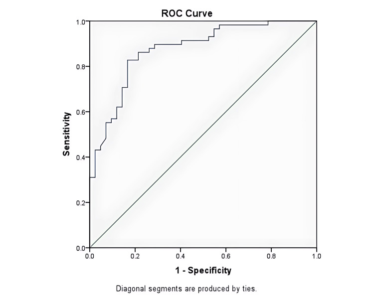
Background and objective Bladder cancer (BC) is a common urothelial neoplasm, with non-muscle invasive forms comprising about 75% of cases and generally having better outcomes than muscle-invasive types. Accurate preoperative grading and staging of BC are essential for appropriate treatment planning. This study investigates the efficacy of computerized tomography (CT) in correlating the morphological features of tumors to predict the histopathological grades of BC. Materials and methods This retrospective cohort involved 100 patients diagnosed with non-muscle invasive BC, who underwent transurethral resection of bladder tumor (TUR-BT) between January 2010 and August 2021. CT imaging, utilizing a 128-slice CT scanner, was employed to measure the tumor height (H) and contact length (CL). The study considered morphometric parameters across axial, coronal, and sagittal planes. Statistical analyses were conducted, comparing radiological findings with histopathological evaluations. Tumor grading was determined according to the 2004/2016 WHO classification. Results Among the 100 patients with primary bladder tumors, 15 were female and 85 were male, with a mean age of 65.28 ± 7.11 years. Furthermore, 58 had high-grade bladder tumors, while 42 had low-grade bladder tumors. Across all planes, high-grade tumors exhibited higher values for the tumor H, CL, and the tumor height-to-contact length (H/CL) ratio compared to low-grade tumors (p<0.05). Notably, the specificity, sensitivity, and diagnostic accuracy of the tumor CL were higher than those of the tumor H and the tumor H/CL ratio. A tumor CL exceeding 19.1mm measured in the axial plane demonstrated 83% sensitivity and specificity for high-grade tumors. Conclusion The measured CL of the tumor in the axial plane on computerized tomography urography has high sensitivity and specificity in detecting high-grade tumors.
膀胱癌(BC)是一种常见的尿路上皮肿瘤,非肌层浸润性形式约占病例的75%,其预后通常比肌层浸润性类型更好。准确的膀胱癌术前分级和分期对于制定合适的治疗方案至关重要。本研究调查计算机断层扫描(CT)在关联肿瘤形态学特征以预测膀胱癌组织病理学分级方面的有效性。
本回顾性队列研究纳入了100例诊断为非肌层浸润性膀胱癌的患者,这些患者在2010年1月至2021年8月期间接受了经尿道膀胱肿瘤切除术(TUR-BT)。使用128层CT扫描仪进行CT成像,测量肿瘤高度(H)和接触长度(CL)。该研究考虑了轴位、冠状位和矢状位平面的形态计量学参数。进行了统计分析,将放射学结果与组织病理学评估进行比较。肿瘤分级根据2004/2016年世界卫生组织分类确定。
在100例原发性膀胱肿瘤患者中,女性15例,男性85例,平均年龄为65.28±7.11岁。此外,58例为高级别膀胱肿瘤,42例为低级别膀胱肿瘤。在所有平面上,与低级别肿瘤相比,高级别肿瘤的肿瘤H、CL以及肿瘤高度与接触长度(H/CL)比值更高(p<0.05)。值得注意的是,肿瘤CL的特异性、敏感性和诊断准确性高于肿瘤H和肿瘤H/CL比值。在轴位平面上测量的肿瘤CL超过19.1mm对高级别肿瘤的敏感性和特异性为83%。
计算机断层扫描尿路造影中轴位平面上测量的肿瘤CL在检测高级别肿瘤方面具有高敏感性和特异性。