AlKhyeli Jaber, Yousif Mohamed Ahmed Mohamed, Abdulgadir Mohanad
Orthopedic Department, Burjeel Medical City, Abu Dhabi, United Arab Emirates.
Orthopedic Department, Burjeel Medical City, Abu Dhabi, United Arab Emirates.
Int J Surg Case Rep. 2024 Sep;122:110105. doi: 10.1016/j.ijscr.2024.110105. Epub 2024 Jul 31.
Due to its ability to provide stable fixation and permit early mobilization, volar plating has become the recommended technique for the surgical stabilization of distal radius fractures. The extensor pollicis longus (EPL) tendon may be injured or ruptured as a result of undetected screw penetration or drill plunging. During surgery, it is critical to detect any potential screw penetration so that it can be corrected.
A 32-year-old woman presented six weeks post-distal radius plating with an inability to extend her left thumb. Clinical examination revealed loss of extension at the interphalangeal joint, stiff wrist, tender point over the dorsal aspect of the wrist, and an intact sensory nerve function.
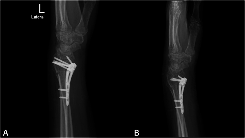
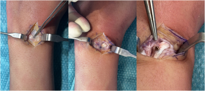
Dynamic ultrasound and magnetic resonance imaging (MRI) both revealed no evidence of tendon rupture or EPL tendon movement. X-rays revealed the distal epiphyseal screws penetrating the far cortex. Intraoperatively, the EPL tendon was found to be impinged by a screw. The tendon was released, tenolysis was performed, and the distal screws were shortened.
In order to assess screw penetration into the far cortex, volar plating for distal radius fractures should be performed using intraoperative imaging views such as lateral, 45-degree supination, 45-degree pronation, dorsal tangential, and skyline views. Timely interventions after distal radius fracture fixation preserve tendon function, and early detection of tendon compromise is essential to preventing additional damage.
由于掌侧钢板能够提供稳定的固定并允许早期活动,它已成为桡骨远端骨折手术固定的推荐技术。未被发现的螺钉穿透或钻孔突进可能导致拇长伸肌腱(EPL)损伤或断裂。在手术过程中,检测任何潜在的螺钉穿透情况并进行纠正至关重要。
一名32岁女性在桡骨远端钢板固定术后六周出现左拇指无法伸展。临床检查发现指间关节伸展功能丧失、手腕僵硬、腕背侧有压痛点,感觉神经功能完好。
动态超声和磁共振成像(MRI)均未显示肌腱断裂或EPL肌腱活动的迹象。X线显示远端骨骺螺钉穿透对侧皮质。术中发现EPL肌腱被一枚螺钉压迫。松解肌腱,进行肌腱松解术,并缩短远端螺钉。
为了评估螺钉是否穿透对侧皮质,桡骨远端骨折掌侧钢板固定应采用术中成像视图,如侧位、45度旋后位、45度旋前位、背侧切线位和天际线位。桡骨远端骨折固定后及时干预可保留肌腱功能,早期发现肌腱损伤对于预防进一步损伤至关重要。