Kling Sabine, Frigelli Matteo, Aydemir M Enes, Tahsini Vahoora, Torres-Netto Emilio A, Kollros Leonard, Hafezi Farhad
Institute for Biomedical Engineering, ITET Department, ETH Zurich, Zurich, Switzerland.
ARTORG Center for Biomedical Engineering Research, University of Bern, Bern, Switzerland.
Commun Med (Lond). 2024 Aug 12;4(1):162. doi: 10.1038/s43856-024-00578-9.
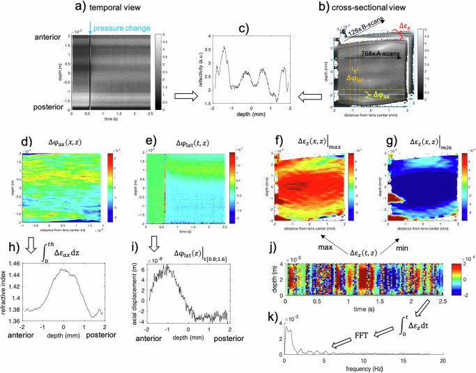
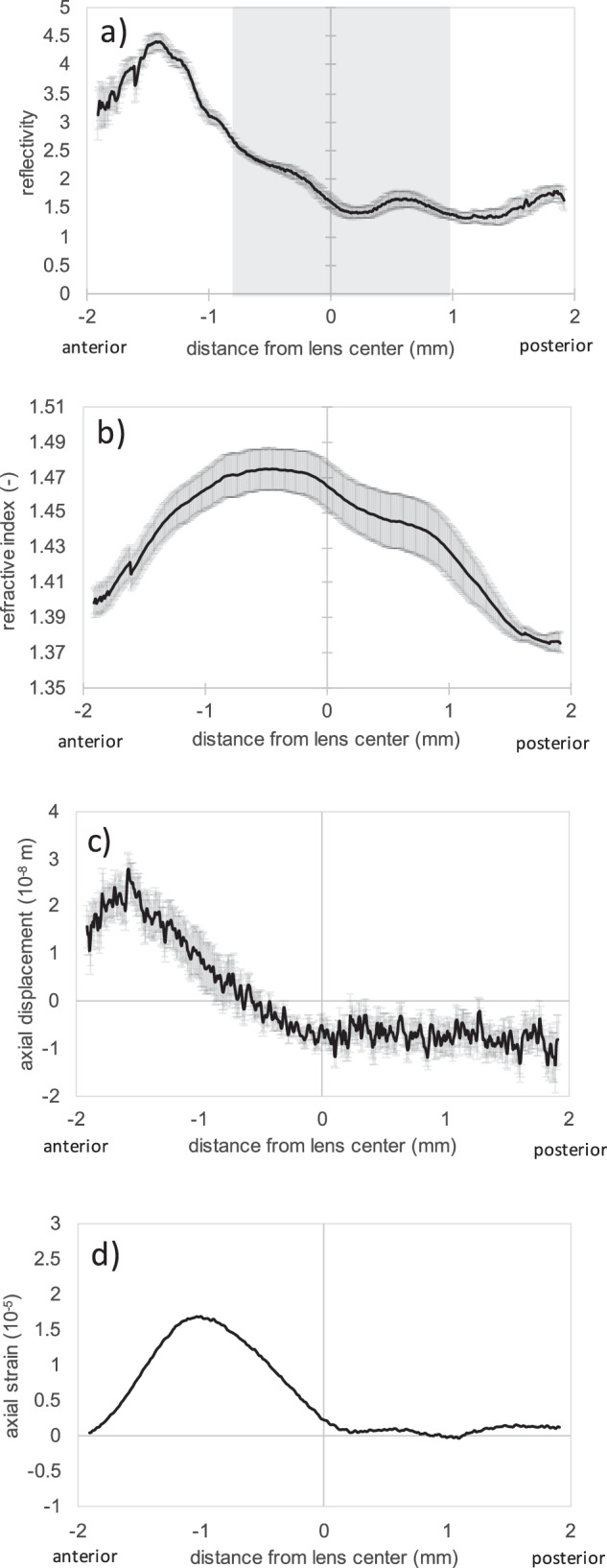
As a key element of ocular accommodation, the inherent mechanical stiffness gradient and the gradient refractive index (GRIN) of the crystalline lens determine its deformability and optical functionality. Quantifying the GRIN profile and deformation characteristics in the lens has the potential to improve the diagnosis and follow-up of lenticular disorders and guide refractive interventions in the future.
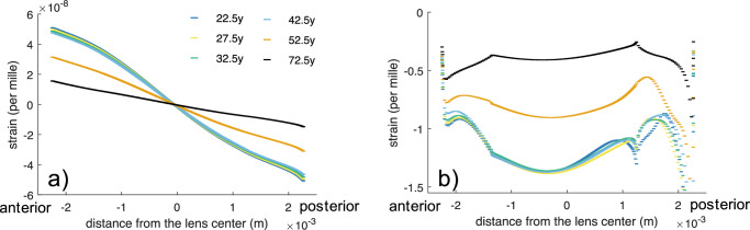
Here, we present a type of optical coherence elastography able to examine the mechanical characteristics of the human crystalline lens and the GRIN distribution in vivo. The concept is demonstrated in a case series of 12 persons through lens displacement and strain measurements in an age-mixed group of human subjects in response to an external (ambient pressure modulation) and an intrinsic (micro-fluctuations of accommodation) mechanical deformation stimulus.
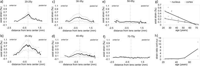
Here we show an excellent agreement between the high-resolution strain map retrieved during steady-state micro-fluctuations and earlier reports on lens stiffness in the cortex and nucleus suggesting a 2.0 to 2.3 times stiffer cortex than the nucleus in young lenses and a 1.0 to 7.0 times stiffer nucleus than the cortex in the old lenses.
Optical coherence tomography is suitable to quantify the internal stiffness and refractive index distribution of the crystalline lens in vivo and thus might contribute to reveal its inner working mechanism. Our methodology provides new routes for ophthalmic pre-surgical examinations and basic research.
作为眼调节的关键要素,晶状体固有的机械刚度梯度和梯度折射率(GRIN)决定了其可变形性和光学功能。量化晶状体中的GRIN分布和变形特征有可能改善晶状体疾病的诊断和随访,并为未来的屈光干预提供指导。
在此,我们展示了一种光学相干弹性成像技术,能够在体内检测人晶状体的机械特性和GRIN分布。通过对一组年龄混合的人类受试者在外部(环境压力调制)和内部(调节的微波动)机械变形刺激下的晶状体位移和应变测量,在12例病例系列中验证了这一概念。
我们展示了在稳态微波动期间获取的高分辨率应变图与早期关于皮质和核中晶状体刚度的报告之间的极佳一致性,表明年轻晶状体中皮质的刚度比核高2.0至2.3倍,而老年晶状体中核的刚度比皮质高1.0至7.0倍。
光学相干断层扫描适用于在体内量化晶状体的内部刚度和折射率分布,因此可能有助于揭示其内部工作机制。我们的方法为眼科术前检查和基础研究提供了新途径。