Save Sight Institute, Discipline of Ophthalmology, Sydney Medical School, The University of Sydney, Sydney, NSW, 2000, Australia.
Centre for Vision Research, Westmead Institute for Medical Research, Faculty of Medicine and Health, Sydney University, Sydney, Westmead, NSW, 2145, Australia.
Sci Rep. 2024 Aug 13;14(1):18752. doi: 10.1038/s41598-024-66068-z.
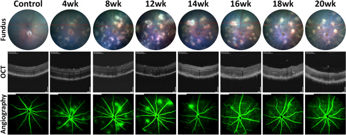
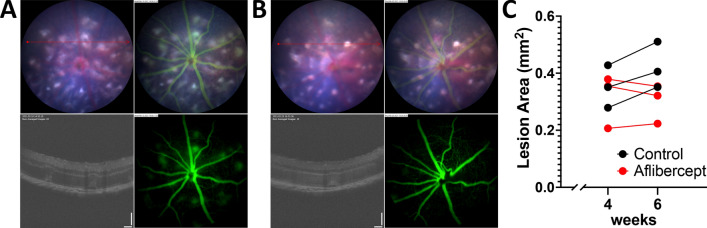
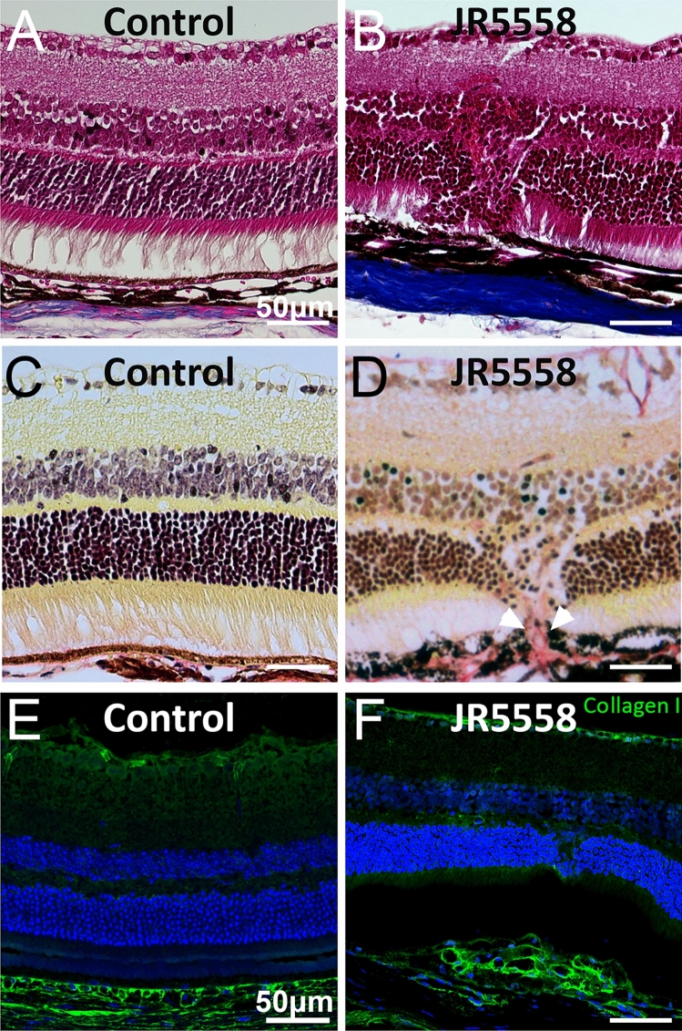
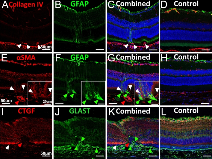
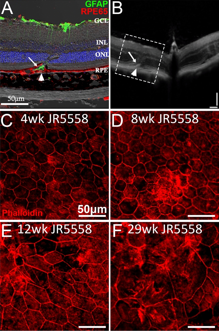
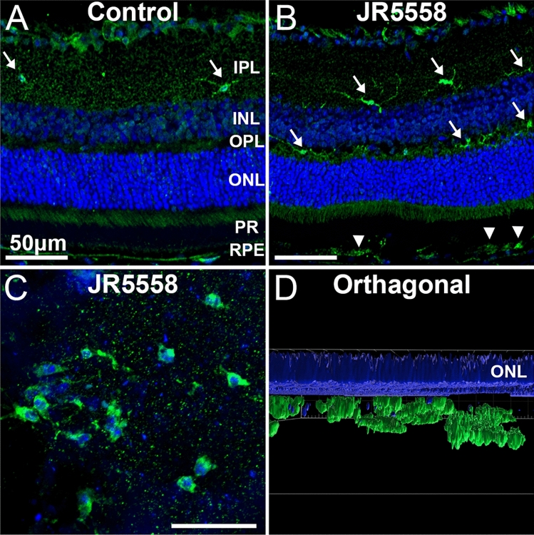
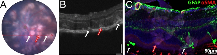
Subretinal fibrosis is a major untreatable cause of poor outcomes in neovascular age-related macular degeneration. Mouse models of subretinal fibrosis all possess a degree of invasiveness and tissue damage not typical of fibrosis progression. This project characterises JR5558 mice as a model to study subretinal fibrosis. Fundus and optical coherence tomography (OCT) imaging was used to non-invasively track lesions. Lesion number and area were quantified with ImageJ. Retinal sections, wholemounts and Western blots were used to characterise alterations. Subretinal lesions expand between 4 and 8 weeks and become established in size and location around 12 weeks. Subretinal lesions were confirmed to be fibrotic, including various cell populations involved in fibrosis development. Müller cell processes extended from superficial retina into subretinal lesions at 8 weeks. Western blotting revealed increases in fibronectin (4 wk and 8 wk, p < 0.001), CTGF (20 wks, p < 0.001), MMP2 (12 wks and 20 wks p < 0.05), αSMA (12 wks and 20 wks p < 0.05) and GFAP (8 wk and 12 wk, p ≤ 0.01), consistent with our immunofluorescence results. Intravitreal injection of Aflibercept reduced subretinal lesion growth. Our study provides evidence JR5558 mice have subretinal fibrotic lesions that grow between 4 and 8 weeks and confirms this line to be a good model to study subretinal fibrosis development and assess treatment options.
视网膜下纤维化是新生血管性年龄相关性黄斑变性不良结局的一个主要不可治疗的原因。视网膜下纤维化的小鼠模型都具有一定程度的侵袭性和组织损伤,这与纤维化进展的特点不典型。本项目将 JR5558 小鼠鉴定为研究视网膜下纤维化的模型。使用眼底和光学相干断层扫描(OCT)成像技术对病变进行非侵入性跟踪。使用 ImageJ 对病变数量和面积进行定量分析。使用视网膜切片、全铺片和 Western blot 对改变进行特征描述。视网膜下病变在 4 至 8 周之间扩大,并在 12 周左右在大小和位置上稳定下来。证实视网膜下病变是纤维化的,包括参与纤维化发展的各种细胞群体。Müller 细胞突起从浅层视网膜延伸到 8 周时的视网膜下病变。Western blot 显示纤维连接蛋白(4 周和 8 周时,p<0.001)、CTGF(20 周时,p<0.001)、MMP2(12 周和 20 周时,p<0.05)、αSMA(12 周和 20 周时,p<0.05)和 GFAP(8 周和 12 周时,p≤0.01)的增加,与免疫荧光结果一致。玻璃体内注射 Aflibercept 可减少视网膜下病变的生长。我们的研究提供了证据表明 JR5558 小鼠具有生长在 4 至 8 周之间的视网膜下纤维性病变,并证实该品系是研究视网膜下纤维化发展和评估治疗选择的良好模型。