Department of Traumatology, University Hospital Zürich, University of Zurich, Zurich, Switzerland.
Department of Orthopaedics, Balgrist University Hospital, Zurich, Switzerland.
BMC Med Imaging. 2024 Aug 13;24(1):213. doi: 10.1186/s12880-024-01395-1.
This study investigated potential use of computed tomography (CT)-based parameters in the lumbar spine as a surrogate for magnetic resonance imaging (MRI)-based findings.
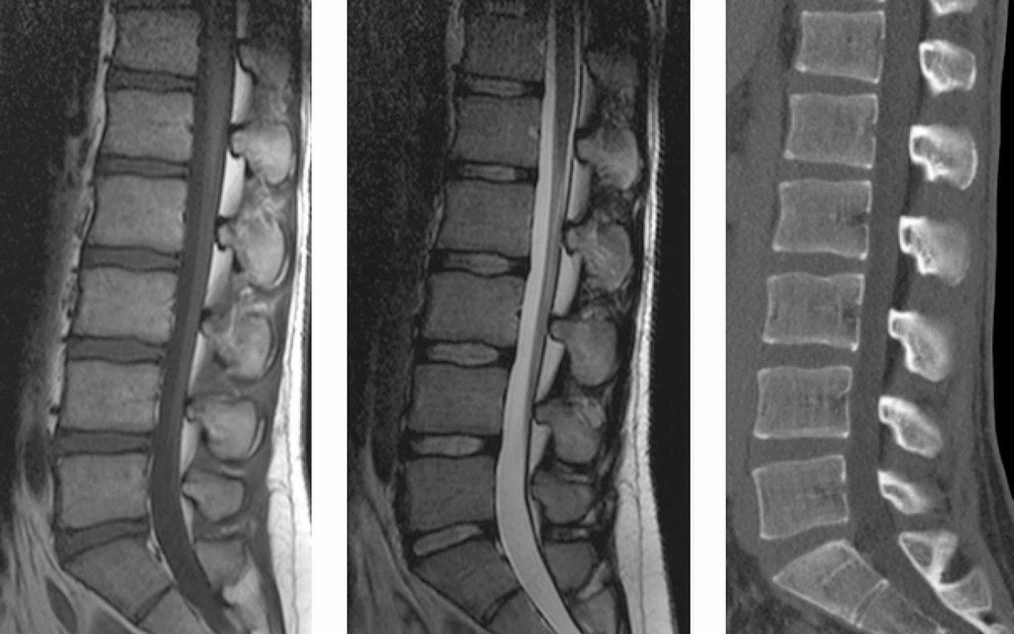
In this retrospective study, all individuals, who had a lumbar spine CT scan and MRI between 2006 and 2012 were reviewed (n = 198). Disc height (DH) and endplate degeneration (ED) were evaluated between Th12/L1-L5/S1. Statistics consisted of Spearman correlation and univariate/multivariable regression (adjusting for age and gender).
The mean CT-DH increased kranio-caudally (8.04 millimeters (mm) at T12/L1, 9.17 mm at L1/2, 10.59 mm at L2/3, 11.34 mm at L3/4, 11.42 mm at L4/5 and 10.47 mm at L5/S1). MRI-ED was observed in 58 (29%) individuals. CT-DH and MRI-DH had strong to very strong correlations (rho 0.781-0.904, p < .001). MRI-DH showed higher absolute values than CT-DH (mean of 1.76 mm). There was a significant association between CT-DH and MRI-ED at L2/3 (p = .006), L3/4 (p = .002), L4/5 (p < .001) and L5/S1 (p < .001). A calculated cut-off point was set at 11 mm.
In the lumbar spine, there is a correlation between disc height on CT and MRI. This can be useful in trauma and emergency cases, where CT is readily available in the lack of an MRI. In addition, in the middle and lower part of the lumbar spine, loss of disc height on CT scans is associated with more pronounced endplate degeneration on MRIs. If the disc height on CT scans is lower than 11 mm, endplate degeneration on MRIs is likely more pronounced.
Level III, a retrospective study.
本研究旨在探讨基于计算机断层扫描(CT)的腰椎参数作为磁共振成像(MRI)基础发现的替代指标的潜在用途。
本回顾性研究纳入了 2006 年至 2012 年间行腰椎 CT 扫描和 MRI 的所有个体(n=198)。在 T12/L1-L5/S1 之间评估椎间盘高度(DH)和终板退变(ED)。统计学分析包括 Spearman 相关分析和单变量/多变量回归(调整年龄和性别因素)。
CT-DH 平均值从颅侧到尾侧逐渐增加(T12/L1 为 8.04 毫米,L1/2 为 9.17 毫米,L2/3 为 10.59 毫米,L3/4 为 11.34 毫米,L4/5 为 11.42 毫米,L5/S1 为 10.47 毫米)。58 例(29%)个体存在 MRI-ED。CT-DH 与 MRI-DH 具有强到非常强的相关性(rho 0.781-0.904,p<.001)。MRI-DH 的绝对值高于 CT-DH(平均 1.76 毫米)。在 L2/3(p=.006)、L3/4(p=.002)、L4/5(p<.001)和 L5/S1(p<.001),CT-DH 与 MRI-ED 之间存在显著相关性。设定了 11 毫米的计算截断值。
在腰椎中,CT 上的椎间盘高度与 MRI 之间存在相关性。在缺乏 MRI 的情况下,CT 可用于创伤和急诊情况,这非常有用。此外,在腰椎的中下部,CT 扫描上的椎间盘高度降低与 MRI 上更明显的终板退变相关。如果 CT 扫描上的椎间盘高度低于 11 毫米,则 MRI 上的终板退变可能更明显。
三级,回顾性研究。