文献检索文档翻译深度研究
Suppr Zotero 插件Zotero 插件
邀请有礼套餐&价格历史记录

新学期,新优惠

限时优惠:9月1日-9月22日

30天高级会员仅需29元

1天体验卡首发特惠仅需5.99元

了解详情
不再提醒
插件&应用
Suppr Zotero 插件Zotero 插件浏览器插件Mac 客户端Windows 客户端微信小程序
高级版
套餐订阅购买积分包
AI 工具
文献检索文档翻译深度研究
关于我们
关于 Suppr公司介绍联系我们用户协议隐私条款
关注我们

Suppr 超能文献

核心技术专利:CN118964589B侵权必究
粤ICP备2023148730 号-1Suppr @ 2025

空间多组学揭示了与人类肝癌癌干细胞特性相关的成纤维细胞亚群。

Spatial multiomics reveals a subpopulation of fibroblasts associated with cancer stemness in human hepatocellular carcinoma.

机构信息

CAS Key Laboratory of Computational Biology, Shanghai Institute of Nutrition and Health, University of Chinese Academy of Sciences, Chinese Academy of Sciences, Shanghai, 200031, People's Republic of China.

Molecular Pathology Laboratory, National Center for Liver Cancer, Eastern Hepatobiliary Surgery Hospital, Shanghai, 201800, People's Republic of China.

出版信息

Genome Med. 2024 Aug 13;16(1):98. doi: 10.1186/s13073-024-01367-8.


DOI:10.1186/s13073-024-01367-8
PMID:39138551
原文链接:https://pmc.ncbi.nlm.nih.gov/articles/PMC11320883/
Abstract

BACKGROUND: Cancer-associated fibroblasts (CAFs) are the prominent cell type in the tumor microenvironment (TME), and CAF subsets have been identified in various tumors. However, how CAFs spatially coordinate other cell populations within the liver TME to promote cancer progression remains unclear. METHODS: 
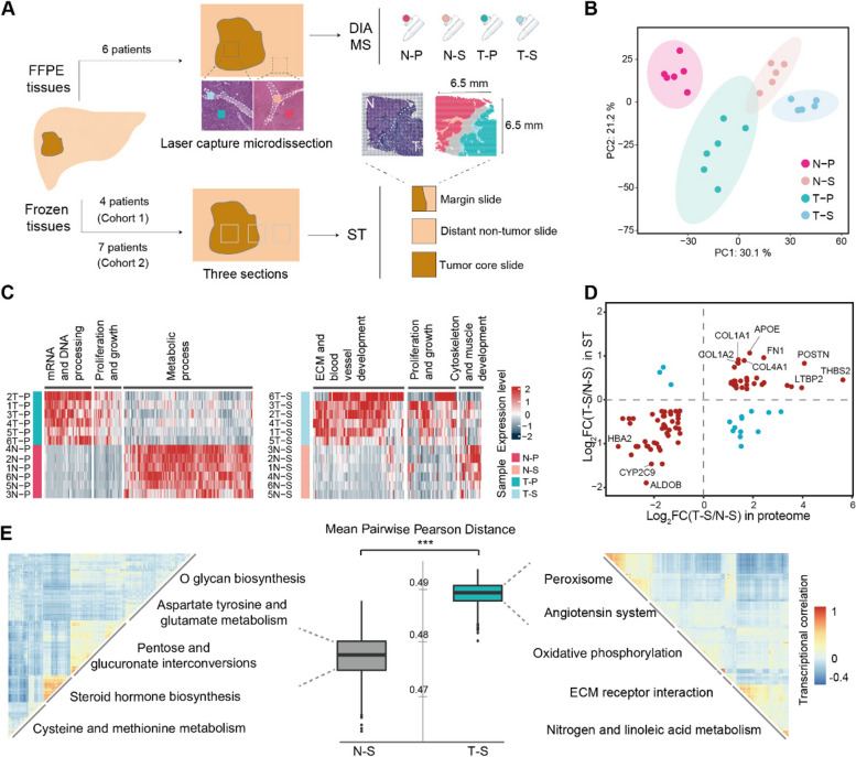
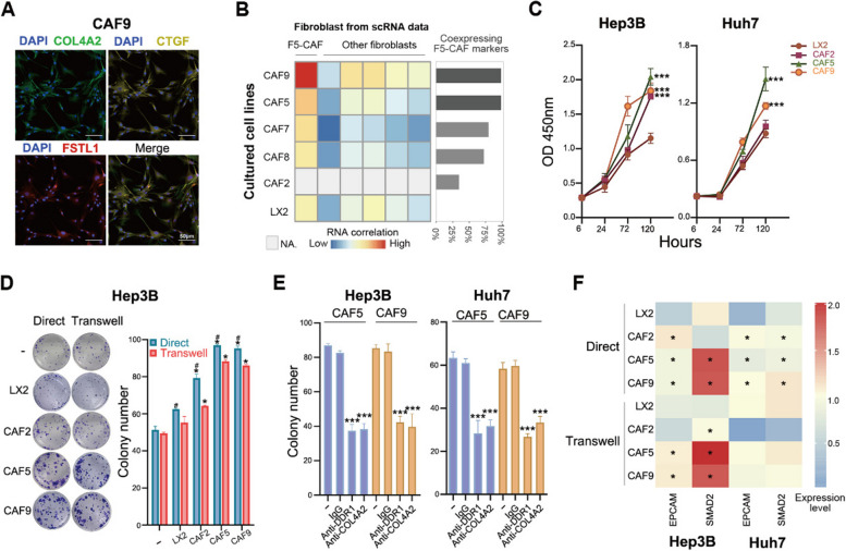
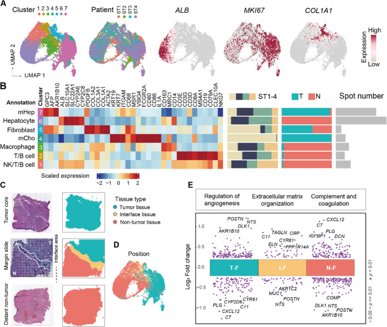
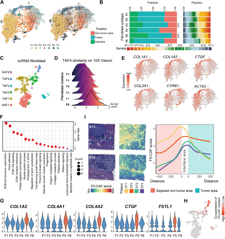
We combined multi-region proteomics (6 patients, 24 samples), 10X Genomics Visium spatial transcriptomics (11 patients, 25 samples), and multiplexed imaging (92 patients, 264 samples) technologies to decipher the expression heterogeneity, functional diversity, spatial distribution, colocalization, and interaction of fibroblasts. The newly identified CAF subpopulation was validated by cells isolated from 5 liver cancer patients and in vitro functional assays. RESULTS: 
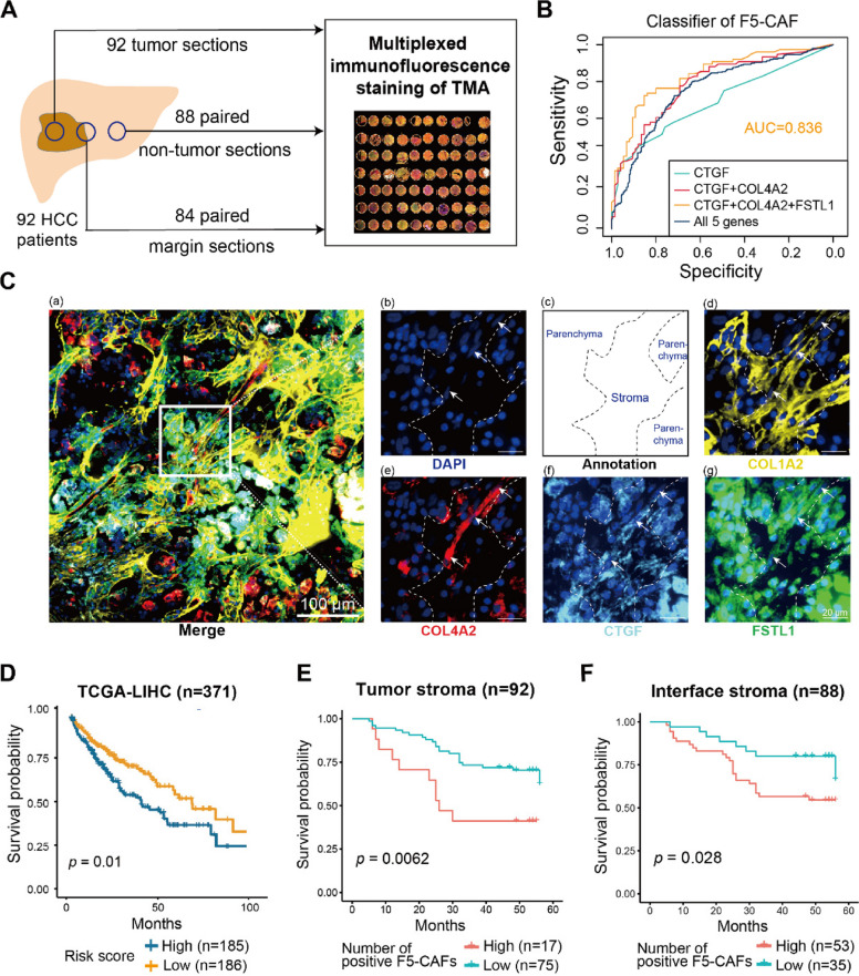
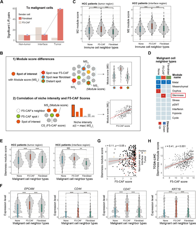
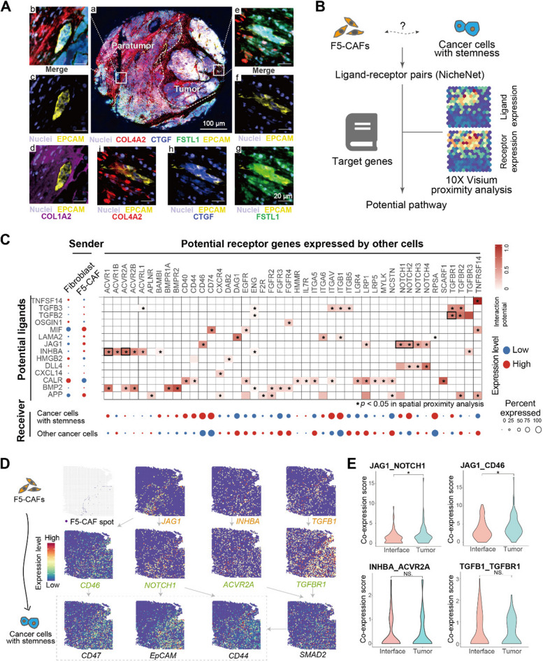
We identified a liver CAF subpopulation, marked by the expression of COL1A2, COL4A1, COL4A2, CTGF, and FSTL1, and named F5-CAF. F5-CAF is preferentially located within and around tumor nests and colocalizes with cancer cells with higher stemness in hepatocellular carcinoma (HCC). Multiplexed staining of 92 patients and the bulk transcriptome of 371 patients demonstrated that the abundance of F5-CAFs in HCC was associated with a worse prognosis. Further in vitro experiments showed that F5-CAFs isolated from liver cancer patients can promote the proliferation and stemness of HCC cells. CONCLUSIONS: We identified a CAF subpopulation F5-CAF in liver cancer, which is associated with cancer stemness and unfavorable prognosis. Our results provide potential mechanisms by which the CAF subset in the TME promotes the development of liver cancer by supporting the survival of cancer stem cells.

摘要

背景:癌症相关成纤维细胞(CAFs)是肿瘤微环境(TME)中的主要细胞类型,并且已经在各种肿瘤中鉴定出 CAF 亚群。然而,CAFs 如何在肝 TME 中与其他细胞群体空间协调以促进癌症进展仍不清楚。

方法:我们结合多区域蛋白质组学(6 名患者,24 个样本)、10X Genomics Visium 空间转录组学(11 名患者,25 个样本)和多重成像(92 名患者,264 个样本)技术,以解析成纤维细胞的表达异质性、功能多样性、空间分布、共定位和相互作用。通过从 5 名肝癌患者分离的细胞和体外功能测定验证了新鉴定的 CAF 亚群。

结果:我们鉴定出一种肝 CAF 亚群,其特征在于 COL1A2、COL4A1、COL4A2、CTGF 和 FSTL1 的表达,并将其命名为 F5-CAF。F5-CAF 优先位于肿瘤巢内和周围,并与肝癌中的具有更高干性的癌细胞共定位。92 名患者的多重染色和 371 名患者的批量转录组学表明,F5-CAF 在 HCC 中的丰度与预后不良相关。进一步的体外实验表明,从肝癌患者中分离的 F5-CAF 可以促进 HCC 细胞的增殖和干性。

结论:我们在肝癌中鉴定出一种 CAF 亚群 F5-CAF,它与癌症干性和不良预后相关。我们的结果提供了潜在的机制,即 TME 中的 CAF 亚群通过支持癌症干细胞的存活来促进肝癌的发展。

https://cdn.ncbi.nlm.nih.gov/pmc/blobs/3bd5/11320883/b8e834158e5f/13073_2024_1367_Fig7_HTML.jpg
https://cdn.ncbi.nlm.nih.gov/pmc/blobs/3bd5/11320883/2991551cd929/13073_2024_1367_Fig1_HTML.jpg
https://cdn.ncbi.nlm.nih.gov/pmc/blobs/3bd5/11320883/c76659523e3d/13073_2024_1367_Fig2_HTML.jpg
https://cdn.ncbi.nlm.nih.gov/pmc/blobs/3bd5/11320883/42cfabaff2a0/13073_2024_1367_Fig3_HTML.jpg
https://cdn.ncbi.nlm.nih.gov/pmc/blobs/3bd5/11320883/ce8237d4b8db/13073_2024_1367_Fig4_HTML.jpg
https://cdn.ncbi.nlm.nih.gov/pmc/blobs/3bd5/11320883/36c149beb5bf/13073_2024_1367_Fig5_HTML.jpg
https://cdn.ncbi.nlm.nih.gov/pmc/blobs/3bd5/11320883/3f81da03c776/13073_2024_1367_Fig6_HTML.jpg
https://cdn.ncbi.nlm.nih.gov/pmc/blobs/3bd5/11320883/b8e834158e5f/13073_2024_1367_Fig7_HTML.jpg

相似文献

[1]
Spatial multiomics reveals a subpopulation of fibroblasts associated with cancer stemness in human hepatocellular carcinoma.

Genome Med. 2024-8-13

[2]
Resolvin D1 prevents epithelial-mesenchymal transition and reduces the stemness features of hepatocellular carcinoma by inhibiting paracrine of cancer-associated fibroblast-derived COMP.

J Exp Clin Cancer Res. 2019-4-18

[3]
Spatial transcriptomics reveals tumor-derived SPP1 induces fibroblast chemotaxis and activation in the hepatocellular carcinoma microenvironment.

J Transl Med. 2024-9-12

[4]
Cancer-associated fibroblasts promote the stemness of CD24 liver cells via paracrine signaling.

J Mol Med (Berl). 2018-12-18

[5]
Prognostic Value of Cancer-Associated Fibroblast-Related Gene Signatures in Hepatocellular Carcinoma.

Front Endocrinol (Lausanne). 2022

[6]
Semaphorin 3C (Sema3C) reshapes stromal microenvironment to promote hepatocellular carcinoma progression.

Signal Transduct Target Ther. 2024-7-3

[7]
FOXQ1/NDRG1 axis exacerbates hepatocellular carcinoma initiation via enhancing crosstalk between fibroblasts and tumor cells.

Cancer Lett. 2017-12-15

[8]
Cancer-Associated Fibroblast-Mediated Cellular Crosstalk Supports Hepatocellular Carcinoma Progression.

Hepatology. 2021-5

[9]
Pancreatic Tumor Microenvironment Factor Promotes Cancer Stemness via SPP1-CD44 Axis.

Gastroenterology. 2021-12

[10]
FSTL1 Secreted by Activated Fibroblasts Promotes Hepatocellular Carcinoma Metastasis and Stemness.

Cancer Res. 2021-11-15

引用本文的文献

[1]
Cancer-associated fibroblasts in hepatocellular carcinoma: origins, heterogeneity, and therapeutic implications.

Front Immunol. 2025-7-18

[2]
Comparative transcriptomic and genomic analysis of tumor cells in the marginal and center regions of tumor nests in human hepatocellular carcinoma.

Front Cell Dev Biol. 2025-7-16

[3]
Ectopic expression of GDF15 in cancer-associated fibroblasts enhances melanoma immunosuppression via the GFRAL/RET cascade.

J Immunother Cancer. 2025-6-24

[4]
Bridging Immune Evasion and Vascular Dynamics for Novel Therapeutic Frontiers in Hepatocellular Carcinoma.

Cancers (Basel). 2025-5-31

[5]
Unsupervised machine learning-based stratification and immune deconvolution of liver hepatocellular carcinoma.

BMC Cancer. 2025-5-10

[6]
Benign non-immune cells in tumor microenvironment.

Front Immunol. 2025-4-3

[7]
infected hepatocellular carcinoma exhibits distinct tumor microenvironment and molecular features.

Front Immunol. 2025-3-17

[8]
Quantifying and interpreting biologically meaningful spatial signatures within tumor microenvironments.

NPJ Precis Oncol. 2025-3-11

[9]
Cancer-associated fibroblasts in hepatocellular carcinoma: heterogeneity, mechanisms and therapeutic targets.

Hepatol Int. 2025-4

[10]
Detection of Cancer Stem Cells from Patient Samples.

Cells. 2025-1-20

本文引用的文献

[1]
Single-cell and spatial transcriptomics reveal POSTN cancer-associated fibroblasts correlated with immune suppression and tumour progression in non-small cell lung cancer.

Clin Transl Med. 2023-12

[2]
Spatial and Single-Cell Transcriptomics Reveal a Cancer-Associated Fibroblast Subset in HNSCC That Restricts Infiltration and Antitumor Activity of CD8+ T Cells.

Cancer Res. 2024-1-16

[3]
Cancer-associated fibroblasts undergoing neoadjuvant chemotherapy suppress rectal cancer revealed by single-cell and spatial transcriptomics.

Cell Rep Med. 2023-10-17

[4]
Pan-cancer spatially resolved single-cell analysis reveals the crosstalk between cancer-associated fibroblasts and tumor microenvironment.

Mol Cancer. 2023-10-13

[5]
Cancer-associated fibroblast classification in single-cell and spatial proteomics data.

Nat Commun. 2023-7-18

[6]
CD36 cancer-associated fibroblasts provide immunosuppressive microenvironment for hepatocellular carcinoma via secretion of macrophage migration inhibitory factor.

Cell Discov. 2023-3-6

[7]
Single-cell RNA sequencing and spatial transcriptomics reveal cancer-associated fibroblasts in glioblastoma with protumoral effects.

J Clin Invest. 2023-3-1

[8]
Identification of a tumour immune barrier in the HCC microenvironment that determines the efficacy of immunotherapy.

J Hepatol. 2023-4

[9]
Colorectal Cancer Metastases in the Liver Establish Immunosuppressive Spatial Networking between Tumor-Associated SPP1+ Macrophages and Fibroblasts.

Clin Cancer Res. 2023-1-4

[10]
Opposing roles of hepatic stellate cell subpopulations in hepatocarcinogenesis.

Nature. 2022-10

文献AI研究员

20分钟写一篇综述,助力文献阅读效率提升50倍

立即体验

用中文搜PubMed

大模型驱动的PubMed中文搜索引擎

马上搜索

推荐工具

医学文档翻译智能文献检索