• 文献检索
  • 文档翻译
  • 深度研究
  • 学术资讯
  • Suppr Zotero 插件Zotero 插件
  • 邀请有礼
  • 套餐&价格
  • 历史记录
应用&插件
Suppr Zotero 插件Zotero 插件浏览器插件Mac 客户端Windows 客户端微信小程序
定价
高级版会员购买积分包购买API积分包
服务
文献检索文档翻译深度研究API 文档MCP 服务
关于我们
关于 Suppr公司介绍联系我们用户协议隐私条款
关注我们

Suppr 超能文献

核心技术专利:CN118964589B侵权必究
粤ICP备2023148730 号-1Suppr @ 2026

文献检索

告别复杂PubMed语法,用中文像聊天一样搜索,搜遍4000万医学文献。AI智能推荐,让科研检索更轻松。

立即免费搜索

文件翻译

保留排版,准确专业,支持PDF/Word/PPT等文件格式,支持 12+语言互译。

免费翻译文档

深度研究

AI帮你快速写综述,25分钟生成高质量综述,智能提取关键信息,辅助科研写作。

立即免费体验

新的毛细血管前性肺动脉高压死亡率预测增量模型。

New Incremental Model for Predicting Mortality in Pre-Capillary Pulmonary Hypertension.

机构信息

Universidade Federal da Paraíba, João Pessoa, PB - Brasil.

Universidade de São Paulo Faculdade de Medicina Hospital das Clínicas Instituto do Coração, São Paulo, SP - Brasil.

出版信息

Arq Bras Cardiol. 2024 Aug 12;121(7):e20230669. doi: 10.36660/abc.20230669. eCollection 2024.

DOI:10.36660/abc.20230669
PMID:39140558
原文链接:https://pmc.ncbi.nlm.nih.gov/articles/PMC11341211/
Abstract

BACKGROUND

In pulmonary hypertension (PH), the identification of easily obtainable prognostic markers associated with right ventricular (RV) dysfunction and survival is needed.

OBJECTIVE

To evaluate the association of red cell distribution width (RDW) with clinical, echocardiographic parameters and survival in patients with pre-capillary PH, with the development of a mortality prediction model.

METHODS

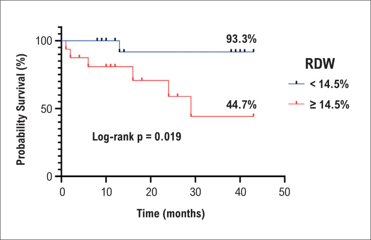
Observational, longitudinal, and prospective study conducted from May 2019 to December 2022. Thirty-four patients with pre-capillary PH underwent two-dimensional echocardiography and complete blood count. A cutoff point of 14.5% was considered to define RDW as altered (≥14.5%) or normal (<14.5%). P values <0.05 were considered significant.

RESULTS

The median RDW was 14.4%. There was a significant difference in peripheral arterial oxygen saturation (SpO2) (p=0.028), RV strain (p=0.047), and pericardial effusion (p=0.002) between the normal and elevated RDW groups. During a median follow-up of 15 months, 20.6% died. Patients with increased RDW had a shorter overall survival (44.7%, log-rank p=0.019), which was a predictor of mortality in univariate Cox regression (HR 8.55, p=0.048). The addition of RV strain <16% and SpO2 ≤93% to the model including RDW alone showed incremental value in predicting mortality (χ2=8.2, p=0.049; χ2=12.4, p=0.041), with increased area under the receiver operating characteristic curve (0.729 vs. 0.837 vs. 0.909) and decreased probability of survival (44.7% vs. 35.6% vs. 25%, log-rank p=0.019).

CONCLUSIONS

RDW provides information on the severity of pre-capillary PH by correlating with echocardiographic parameters of RV dysfunction and mortality, which is best predicted by a model including RDW, RV strain and SpO2.

摘要

背景

在肺动脉高压(PH)中,需要确定与右心室(RV)功能障碍和生存相关的易于获得的预后标志物。

目的

评估红细胞分布宽度(RDW)与毛细血管前 PH 患者的临床、超声心动图参数和生存的相关性,并建立死亡率预测模型。

方法

观察性、纵向和前瞻性研究,于 2019 年 5 月至 2022 年 12 月进行。34 例毛细血管前 PH 患者接受二维超声心动图和全血细胞计数检查。将 RDW 定义为改变(≥14.5%)或正常(<14.5%)的截断点为 14.5%。p 值<0.05 被认为具有统计学意义。

结果

RDW 的中位数为 14.4%。RDW 正常和升高组之间,外周动脉血氧饱和度(SpO2)(p=0.028)、RV 应变(p=0.047)和心包积液(p=0.002)有显著差异。在中位随访 15 个月期间,20.6%的患者死亡。RDW 升高的患者总生存率更短(44.7%,log-rank p=0.019),这是单因素 Cox 回归中死亡的预测因素(HR 8.55,p=0.048)。将 RV 应变<16%和 SpO2≤93%加入到仅包括 RDW 的模型中,预测死亡率具有增量价值(χ2=8.2,p=0.049;χ2=12.4,p=0.041),ROC 曲线下面积增加(0.729 比 0.837 比 0.909),生存率降低(44.7%比 35.6%比 25%,log-rank p=0.019)。

结论

RDW 通过与 RV 功能障碍和死亡率的超声心动图参数相关,提供了毛细血管前 PH 严重程度的信息,通过包括 RDW、RV 应变和 SpO2 的模型可以最好地预测。

https://cdn.ncbi.nlm.nih.gov/pmc/blobs/b6a9/11341211/5a11105eb7a1/0066-782X-abc-121-07-e20230669-gf05-en.jpg
https://cdn.ncbi.nlm.nih.gov/pmc/blobs/b6a9/11341211/9f0cb7e3a774/0066-782X-abc-121-07-e20230669-gf06.jpg
https://cdn.ncbi.nlm.nih.gov/pmc/blobs/b6a9/11341211/8f428664589e/0066-782X-abc-121-07-e20230669-gf01.jpg
https://cdn.ncbi.nlm.nih.gov/pmc/blobs/b6a9/11341211/6db7423d62ef/0066-782X-abc-121-07-e20230669-gf02.jpg
https://cdn.ncbi.nlm.nih.gov/pmc/blobs/b6a9/11341211/06ae33e4d409/0066-782X-abc-121-07-e20230669-gf03.jpg
https://cdn.ncbi.nlm.nih.gov/pmc/blobs/b6a9/11341211/9a3f48ed95d2/0066-782X-abc-121-07-e20230669-gf04.jpg
https://cdn.ncbi.nlm.nih.gov/pmc/blobs/b6a9/11341211/5a8d38141b53/0066-782X-abc-121-07-e20230669-gf05.jpg
https://cdn.ncbi.nlm.nih.gov/pmc/blobs/b6a9/11341211/b0aa48e8f554/0066-782X-abc-121-07-e20230669-gf06-en.jpg
https://cdn.ncbi.nlm.nih.gov/pmc/blobs/b6a9/11341211/46ccbbd33d43/0066-782X-abc-121-07-e20230669-gf01-en.jpg
https://cdn.ncbi.nlm.nih.gov/pmc/blobs/b6a9/11341211/3cd257f84514/0066-782X-abc-121-07-e20230669-gf02-en.jpg
https://cdn.ncbi.nlm.nih.gov/pmc/blobs/b6a9/11341211/522f4a5a1908/0066-782X-abc-121-07-e20230669-gf03-en.jpg
https://cdn.ncbi.nlm.nih.gov/pmc/blobs/b6a9/11341211/58815fe6294e/0066-782X-abc-121-07-e20230669-gf04-en.jpg
https://cdn.ncbi.nlm.nih.gov/pmc/blobs/b6a9/11341211/5a11105eb7a1/0066-782X-abc-121-07-e20230669-gf05-en.jpg

相似文献

1
New Incremental Model for Predicting Mortality in Pre-Capillary Pulmonary Hypertension.新的毛细血管前性肺动脉高压死亡率预测增量模型。
Arq Bras Cardiol. 2024 Aug 12;121(7):e20230669. doi: 10.36660/abc.20230669. eCollection 2024.
2
Three-dimensional echocardiography and 2D-3D speckle-tracking imaging in chronic pulmonary hypertension: diagnostic accuracy in detecting hemodynamic signs of right ventricular (RV) failure.三维超声心动图和二维-三维斑点追踪成像在慢性肺动脉高压中的应用:检测右心室(RV)衰竭血流动力学征象的诊断准确性
J Am Heart Assoc. 2015 Mar 19;4(3):e001584. doi: 10.1161/JAHA.114.001584.
3
Prognostic value of right ventricular free wall strain in pulmonary hypertension patients with pseudo-normalized tricuspid annular plane systolic excursion values.三尖瓣环平面收缩期位移值假性正常化的肺动脉高压患者右心室游离壁应变的预后价值
Int J Cardiovasc Imaging. 2016 Jun;32(6):905-12. doi: 10.1007/s10554-016-0862-8. Epub 2016 Mar 1.
4
Outcome prediction by quantitative right ventricular function assessment in 575 subjects evaluated for pulmonary hypertension.575 例肺动脉高压评估患者的定量右心功能评估的预后预测。
Circ Cardiovasc Imaging. 2013 Sep;6(5):711-21. doi: 10.1161/CIRCIMAGING.113.000640. Epub 2013 Jun 27.
5
Prognostic Value of Right Ventricular Longitudinal Strain in Patients With COVID-19.新冠肺炎患者右心室纵向应变的预后价值。
JACC Cardiovasc Imaging. 2020 Nov;13(11):2287-2299. doi: 10.1016/j.jcmg.2020.04.014. Epub 2020 Apr 28.
6
The prognostic value of right ventricular outflow tract velocity time integral in patients with pulmonary hypertension.右心室流出道速度时间积分对肺动脉高压患者的预后价值。
ESC Heart Fail. 2024 Oct;11(5):3332-3340. doi: 10.1002/ehf2.14934. Epub 2024 Jul 3.
7
Efficacy of right ventricular free-wall longitudinal speckle-tracking strain for predicting long-term outcome in patients with pulmonary hypertension.右心室游离壁纵向斑点追踪应变预测肺动脉高压患者长期预后的效果。
Circ J. 2013;77(3):756-63. doi: 10.1253/circj.cj-12-1083. Epub 2012 Dec 7.
8
Assessment of right ventricular longitudinal strain by 2D speckle tracking imaging compared with RV function and hemodynamics in pulmonary hypertension.二维斑点追踪成像评估右心室纵向应变与肺动脉高压患者右心室功能及血流动力学的比较
Int J Cardiovasc Imaging. 2017 Nov;33(11):1737-1748. doi: 10.1007/s10554-017-1182-3. Epub 2017 May 29.
9
Unbiased Phenome-Wide Association Studies of Red Cell Distribution Width Identifies Key Associations with Pulmonary Hypertension.红细胞分布宽度的无偏phenome-wide 关联研究鉴定出与肺动脉高压的关键关联。
Ann Am Thorac Soc. 2019 May;16(5):589-598. doi: 10.1513/AnnalsATS.201809-594OC.
10
Red Blood Cells Distribution Width as a Potential Prognostic Biomarker in Patients With Pulmonary Arterial Hypertension and Chronic Thromboembolic Pulmonary Hypertension.红细胞分布宽度作为肺动脉高压和慢性血栓栓塞性肺动脉高压患者潜在的预后生物标志物
Heart Lung Circ. 2018 Jul;27(7):842-848. doi: 10.1016/j.hlc.2017.08.007. Epub 2017 Aug 24.

引用本文的文献

1
Serum proteomics identify biomarkers and pathogenesis of portopulmonary hypertension diagnosed based on echocardiography.血清蛋白质组学鉴定基于超声心动图诊断的门肺高压的生物标志物和发病机制。
Sci Rep. 2025 Jul 1;15(1):21075. doi: 10.1038/s41598-025-08376-6.

本文引用的文献

1
2022 ESC/ERS Guidelines for the diagnosis and treatment of pulmonary hypertension.2022年欧洲心脏病学会/欧洲呼吸学会肺动脉高压诊断和治疗指南。
Eur Heart J. 2022 Oct 11;43(38):3618-3731. doi: 10.1093/eurheartj/ehac237.
2
Prognostic impact of red blood cell distribution width in pulmonary hypertension patients: A systematic review and meta-analysis.红细胞分布宽度对肺动脉高压患者的预后影响:一项系统评价和荟萃分析。
Medicine (Baltimore). 2020 Apr;99(16):e19089. doi: 10.1097/MD.0000000000019089.
3
Red Blood Cell Distribution Width Predicts Pulmonary Hypertension Secondary to Chronic Obstructive Pulmonary Disease.
红细胞分布宽度预测慢性阻塞性肺疾病相关肺动脉高压。
Can Respir J. 2019 Jul 11;2019:3853454. doi: 10.1155/2019/3853454. eCollection 2019.
4
Red Cell Distribution Width and Platelet Count as Biomarkers of Pulmonary Arterial Hypertension in Patients with Connective Tissue Disorders.红细胞分布宽度和血小板计数作为结缔组织疾病患者肺动脉高压的生物标志物。
Dis Markers. 2019 Jun 2;2019:4981982. doi: 10.1155/2019/4981982. eCollection 2019.
5
Use of red cell distribution width in a population at high risk for pulmonary hypertension.红细胞分布宽度在肺动脉高压高危人群中的应用。
Respir Med. 2019 Apr;150:131-135. doi: 10.1016/j.rmed.2019.03.003. Epub 2019 Mar 16.
6
Usefulness of Red Cell Width Distribution (RDW) in the Assessment of Children with Pulmonary Arterial Hypertension (PAH).红细胞分布宽度(RDW)在评估儿童肺动脉高压(PAH)中的应用价值。
Pediatr Cardiol. 2019 Apr;40(4):820-826. doi: 10.1007/s00246-019-02077-4. Epub 2019 Mar 4.
7
Haemodynamic definitions and updated clinical classification of pulmonary hypertension.血流动力学定义和肺动脉高压的最新临床分类。
Eur Respir J. 2019 Jan 24;53(1). doi: 10.1183/13993003.01913-2018. Print 2019 Jan.
8
Guidelines for Performing a Comprehensive Transthoracic Echocardiographic Examination in Adults: Recommendations from the American Society of Echocardiography.成人经胸超声心动图全面检查操作指南:美国超声心动图学会的建议
J Am Soc Echocardiogr. 2019 Jan;32(1):1-64. doi: 10.1016/j.echo.2018.06.004. Epub 2018 Oct 1.
9
Red blood cell distribution width as a potential predictor of survival of pulmonary arterial hypertension associated with primary Sjogren's syndrome: a retrospective cohort study.红细胞分布宽度作为原发性干燥综合征相关肺动脉高压患者生存的潜在预测指标:一项回顾性队列研究。
Clin Rheumatol. 2019 Feb;38(2):477-485. doi: 10.1007/s10067-018-4281-1. Epub 2018 Sep 14.
10
Relevance of the TAPSE/PASP ratio in pulmonary arterial hypertension.三尖瓣环收缩期位移与肺动脉收缩压比值在肺动脉高压中的相关性。
Int J Cardiol. 2018 Sep 1;266:229-235. doi: 10.1016/j.ijcard.2018.01.053.