Department of Ophthalmology, The Second Affiliated Hospital, Jiangxi Medical College, Nanchang University, Nanchang, 330006, Jiangxi, China.
Huankui Academy, Nanchang University, Nanchang, 330006, Jiangxi, China.
Sci Rep. 2024 Aug 15;14(1):18935. doi: 10.1038/s41598-024-69493-2.
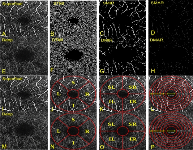
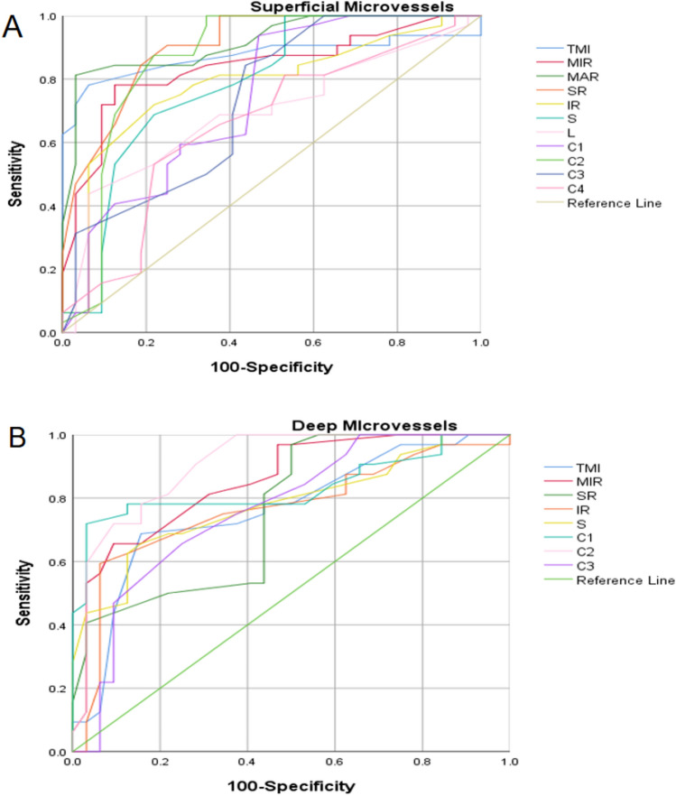
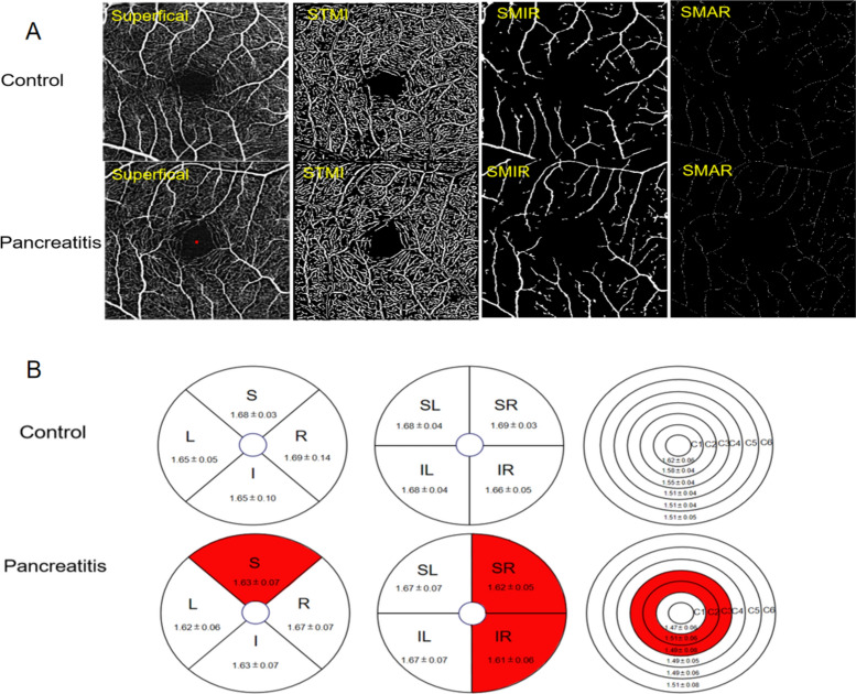
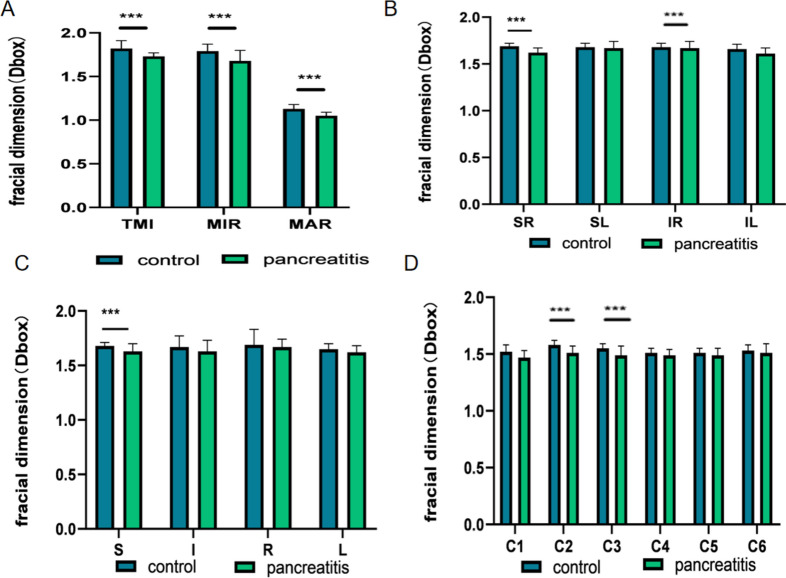
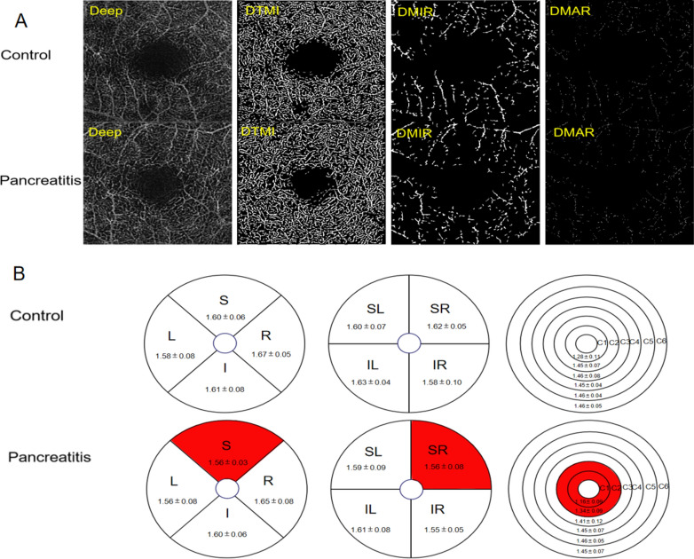
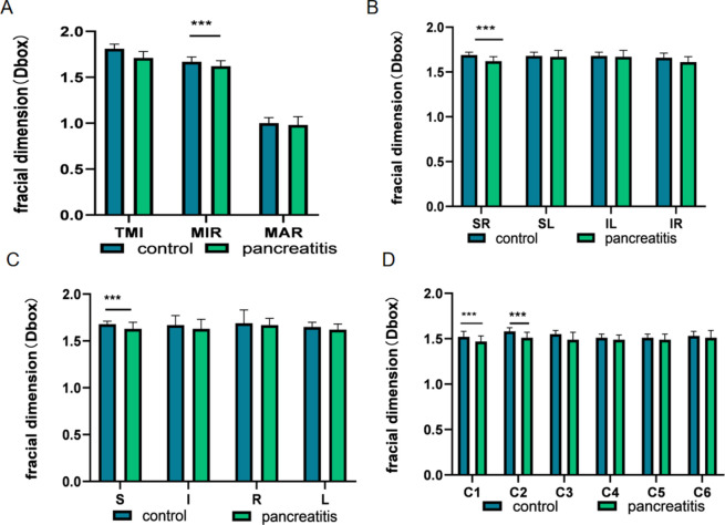
Acute pancreatitis, a common exocrine inflammatory disease affecting the pancreas, is characterized by intense abdominal pain and multiple organ dysfunction. However, the alterations in retinal blood vessels among individuals with acute pancreatitis remain poorly understood. This study employed optical coherence tomography angiography (OCTA) to examine the superficial and deep retinal blood vessels in patients with pancreatitis. Sixteen patients diagnosed with pancreatitis (32 eyes) and 16 healthy controls (32 eyes) were recruited from the First Affiliated Hospital of Nanchang University for participation in the study. Various ophthalmic parameters, such as visual acuity, intraocular pressure, and OCTA image for retina consisting of the superficial retinal layer (SRL) and the deep retinal layer (DRL), were recorded for each eye. The study observed the superficial and deep retinal microvascular ring (MIR), macrovascular ring (MAR), and total microvessels (TMI) were observed. Changes in retinal vascular density in the macula through annular partitioning (C1-C6), hemispheric quadrant partitioning (SR, SL, IL, and IR), and early diabetic retinopathy treatment studies (ETDRS) partitioning methods (R, S, L, and I). Correlation analysis was employed to investigate the relationship between retinal capillary density and clinical indicators. Our study revealed that in the superficial retinal layer, the vascular density of TMI, MIR, MAR, SR, IR, S, C2, C3 regions were significantly decreased in patients group compared with the normal group. For the deep retinal layer, the vascular density of MIR, SR, S, C1, C2 regions also reduced in patient group. The ROC analysis demonstrated that OCTA possesses significant diagnostic performance for pancreatitis. In conclusion, patients with pancreatitis may have retinal microvascular dysfunction, and OCTA can be a valuable tool for detecting alterations in ocular microcirculation in pancreatitis patients in clinical practice.
急性胰腺炎是一种常见的胰腺外分泌炎症性疾病,其特征为剧烈腹痛和多器官功能障碍。然而,急性胰腺炎患者视网膜血管的改变仍知之甚少。本研究采用光学相干断层血管造影(OCTA)检查胰腺炎患者的视网膜浅层和深层血管。从南昌大学第一附属医院招募了 16 名确诊为胰腺炎的患者(32 只眼)和 16 名健康对照者(32 只眼)参与本研究。记录了每只眼的视力、眼压和包括视网膜浅层(SRL)和深层(DRL)在内的 OCTA 视网膜图像等各种眼科参数。观察了视网膜浅层和深层微血管环(MIR)、大血管环(MAR)和总微血管(TMI)。通过环形分区(C1-C6)、半球象限分区(SR、SL、IL 和 IR)和早期糖尿病视网膜病变治疗研究(ETDRS)分区方法(R、S、L 和 I)观察黄斑区视网膜血管密度的变化。采用相关性分析研究视网膜毛细血管密度与临床指标的关系。我们的研究表明,在视网膜浅层,与正常组相比,患者组的 TMI、MIR、MAR、SR、IR、S、C2 和 C3 区域的血管密度显著降低。在深层视网膜,MIR、SR、S、C1 和 C2 区域的血管密度也降低。ROC 分析表明,OCTA 对胰腺炎具有显著的诊断性能。总之,胰腺炎患者可能存在视网膜微血管功能障碍,OCTA 可作为临床检测胰腺炎患者眼部微循环改变的一种有价值的工具。