Thatcher Kelly Lynn, Nielsen Karen Emily, Sandler Evan Blake, Daliet Oliver John, Iddings Jennifer Ann, Field-Fote Edelle Carmen
Shepherd Center.
Georgia State University.
Res Sq. 2024 Aug 7:rs.3.rs-4719031. doi: 10.21203/rs.3.rs-4719031/v1.
There is growing interest in use of transcutaneous spinal stimulation (TSS) for people with neurologic conditions both to augment volitional control (by facilitating motoneuron excitability), and to decrease spasticity (by activating inhibitory networks). Various electrode montages are used during TSS, with little understanding of how electrode position influences spinal circuit activation. We sought to identify the thoracolumbar electrode montage associated with the most robust activation of spinal circuits by comparing posterior root-muscle reflexes (PRM reflexes) elicited by 6 montages. Additionally, we assessed tolerability of the stimulation during PRM reflex testing.
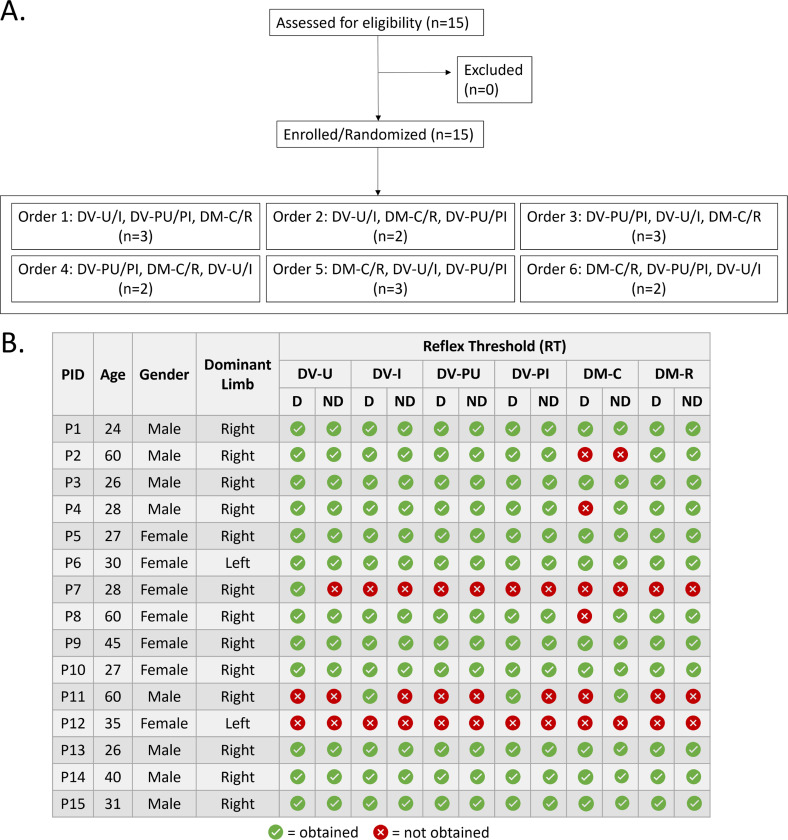
Fifteen adults with intact neurological systems participated in this randomized crossover study. PRM reflexes were evoked transcutaneously using electrode montages with dorsal-ventral (DV) or dorsal-midline (DM) current flow. DV montages included: [1] cathode over T11/T12, anodes over iliac crests (DV-I), [2] cathode over T11/T12, anodes over umbilicus (DV-U), [3] dual paraspinal cathodes at T11/12, anodes over iliac crests (DV-PI), and [4] dual paraspinal cathodes at T11/12, anodes over umbilicus (DV-PU). DM montages included: [5] cathode over T11/12, anode 5cm caudal (DM-C), and [6] cathode over T11/12, anode 5cm rostral (DM-R). PRM reflex recruitment curves were obtained in the soleus muscle of both lower extremities.
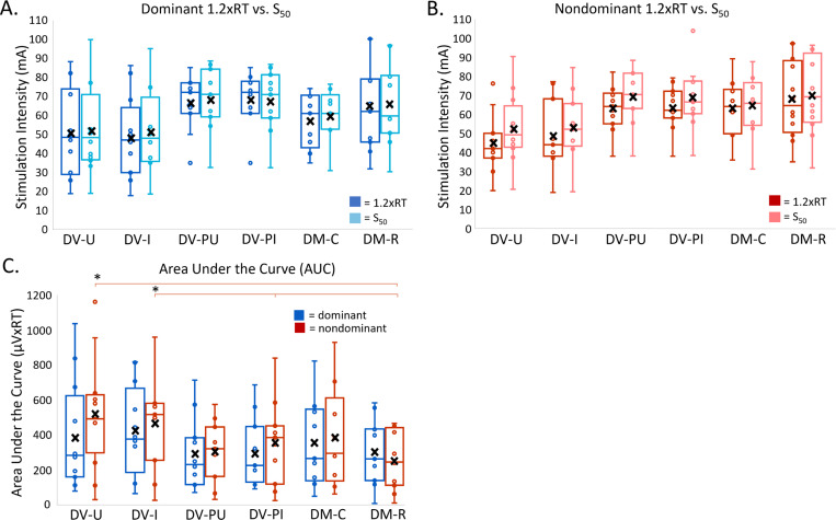
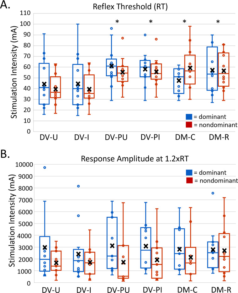
DV-U and DV-I montages elicited bilateral reflexes with lower reflex thresholds and larger recruitment curve area than other montages. There were no differences in response amplitude at 120% of RT(1.2xRT) or tolerability among montages.
Differences in spinal circuit recruitment are reflected in the response amplitude of the PRM reflexes. DV-I and DV-U montages were associated with lower reflex thresholds, indicating that motor responses can be evoked with lower stimulation intensity. DV-I and DV-U montages therefore have the potential for lower and more tolerable interventional stimulation intensities. Our findings optimize electrode placement for interventional TSS and PRM reflex assessments.
经皮脊髓刺激(TSS)在神经系统疾病患者中的应用越来越受到关注,其目的一是增强意志控制(通过促进运动神经元兴奋性),二是减轻痉挛(通过激活抑制性网络)。TSS过程中使用了各种电极组合,但对于电极位置如何影响脊髓回路激活了解甚少。我们试图通过比较6种组合引发的后根 - 肌肉反射(PRM反射),来确定与脊髓回路最强烈激活相关的胸腰段电极组合。此外,我们在PRM反射测试期间评估了刺激的耐受性。
15名神经系统完整的成年人参与了这项随机交叉研究。使用背腹(DV)或背中线(DM)电流流动的电极组合经皮诱发PRM反射。DV组合包括:[1] T11/T12处阴极,髂嵴处阳极(DV - I),[2] T11/T12处阴极,脐部阳极(DV - U),[3] T11/12处双侧椎旁阴极,髂嵴处阳极(DV - PI),以及[4] T11/12处双侧椎旁阴极,脐部阳极(DV - PU)。DM组合包括:[5] T11/12处阴极,尾侧5cm处阳极(DM - C),以及[6] T11/12处阴极,头侧5cm处阳极(DM - R)。在双下肢比目鱼肌中获得PRM反射募集曲线。
DV - U和DV - I组合引发的双侧反射具有比其他组合更低的反射阈值和更大的募集曲线面积。各组合在RT的120%(1.2xRT)时的反应幅度或耐受性方面没有差异。
脊髓回路募集的差异反映在PRM反射的反应幅度中。DV - I和DV - U组合与较低的反射阈值相关,表明可以用较低的刺激强度诱发运动反应。因此,DV - I和DV - U组合有可能采用更低且更耐受的介入刺激强度。我们的研究结果优化了介入性TSS和PRM反射评估的电极放置。