Albanese Giuseppe Maria, Gharbiya Magda, Visioli Giacomo, Panigutti Massimiliano, Margarella Andrea, Romano Enrico, Mastrogiuseppe Elvia, Sepe-Monti Micaela, Bruno Giuseppe, D'Antonio Fabrizia
Department of Sense Organs, Sapienza University of Rome, 155, Viale del Policlinico, Rome, 00161, Italy.
Department of Human Neurosciences, Sapienza University of Rome, Rome, 00185, Italy.
Neurol Sci. 2025 Jan;46(1):185-194. doi: 10.1007/s10072-024-07683-6. Epub 2024 Aug 17.
To explore retinal changes in patients with Dementia with Lewy Bodies (DLB) using Spectral Domain-Optical Coherence Tomography (SD-OCT) and Optical Coherence Tomography Angiography (OCTA), aiming to identify potential biomarkers for diagnosis and monitoring.
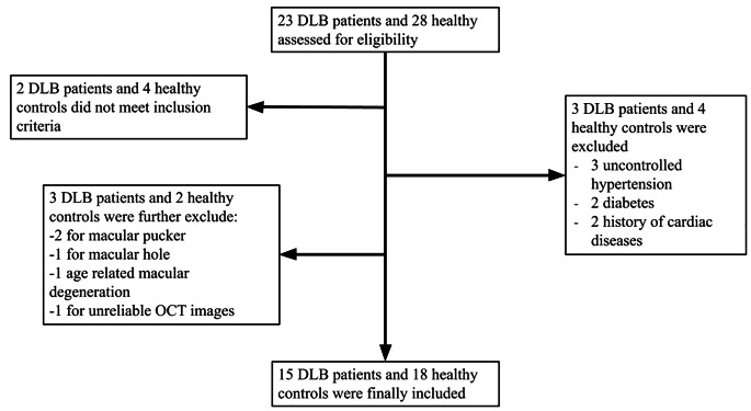
A cross-sectional study analyzed 15 DLB patients and 18 matched controls. Participants underwent physical, neurological, neuropsychological, and ophthalmological evaluations, including SD-OCT and OCTA. Logistic regression, adjusted for age, sex, and inter-eye correlation, was employed to identify retinal alterations in patients affected by DLB.
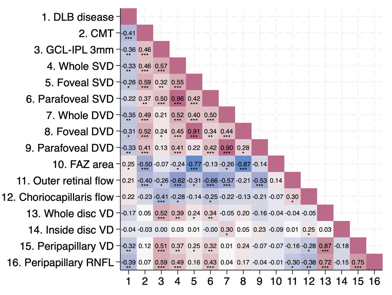
OCTA revealed that DLB is associated with reduced superficial and deep vessel densities (SVD and DVD) in the macula (p < 0.01), as well as decreased peripapillary vessel density (ppVD, p < 0.01). SD-OCT parameters showed correlations with DLB, including reduced central macular thickness (CMT, p < 0.001) and thinning of the ganglion cell layer-inner plexiform layer (GCL-IPL, p < 0.01). Logistic regression (R²=0.26) identified reduced ppVD as a significant predictor of DLB (p = 0.030).
Impairments in retinal capillaries, especially lower ppVD, might mirror cerebral hypoperfusion in DLB, potentially due to reduced Vascular Endothelial Growth Factor (VEGF) levels and increased α-synuclein. Further investigations are warranted to confirm the causal relationship between these observations, disease severity, and progression, as well as their potential role as biomarkers for DLB.
使用光谱域光学相干断层扫描(SD-OCT)和光学相干断层扫描血管造影(OCTA)探索路易体痴呆(DLB)患者的视网膜变化,旨在确定用于诊断和监测的潜在生物标志物。
一项横断面研究分析了15例DLB患者和18例匹配的对照。参与者接受了体格、神经、神经心理和眼科评估,包括SD-OCT和OCTA。采用经年龄、性别和眼间相关性调整的逻辑回归来确定DLB患者的视网膜改变。
OCTA显示,DLB与黄斑区浅表和深部血管密度(SVD和DVD)降低相关(p < 0.01),以及视盘周围血管密度降低(ppVD,p < 0.01)。SD-OCT参数显示与DLB相关,包括中心黄斑厚度降低(CMT,p < 0.001)和神经节细胞层-内丛状层变薄(GCL-IPL,p < 0.01)。逻辑回归(R² = 0.26)确定降低的ppVD是DLB的显著预测指标(p = 0.030)。
视网膜毛细血管损伤,尤其是较低的ppVD,可能反映了DLB中的脑灌注不足,这可能是由于血管内皮生长因子(VEGF)水平降低和α-突触核蛋白增加所致。有必要进行进一步研究以确认这些观察结果、疾病严重程度和进展之间的因果关系,以及它们作为DLB生物标志物的潜在作用。