Akaike Natsuki, Okawa Masakazu, Ishii Akira, Kikuchi Takayuki, Yamao Yukihiro, Abekura Yu, Tsuji Hirofumi, Akiyama Ryo, Matsukawa So, Arakawa Yoshiki
Department of Neurosurgery, Kyoto University School of Medicine, Shogoin Kawahara-Cho Sakyo-ku, Kyoto, Japan.
J Neurosurg Case Lessons. 2024 Aug 19;8(8). doi: 10.3171/CASE2457.
Spinal epidural arteriovenous fistulas (SEAVFs) with intraosseous shunts are rare, and their underlying pathophysiological mechanisms remain unclear.
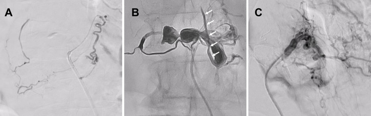
A female in her 70s presented with rapidly progressive weakness in both lower extremities and urinary retention. Lumbar spine magnetic resonance imaging revealed spinal cord edema and flow voids due to venous dilation and compression fractures of the L1 and L2 vertebral bodies. Spinal angiography revealed ventral and dorsal somatic branches of the lumbar arteries at L1 and L2 flowing into the shunt. High-resolution cone-beam computed tomography revealed a shunt within the compression-fractured vertebral body bone of L2. The intravertebral shunt blood flowed into the ventral epidural venous plexus (VEVP) and returned into the perimedullary vein (PMV). Transarterial embolization was performed using N-butyl cyanoacrylate and Onyx-18 for feeder L1 and feeder L2, respectively. Onyx-18 was injected from the VEVP into the PMV, and complete occlusion of the shunt was achieved. The patient showed symptomatic improvement postoperatively.
Vertebral compression fractures are common but rarely associated with SEAVFs. https://thejns.org/doi/10.3171/CASE2457.
伴有骨内分流的脊髓硬膜外动静脉瘘(SEAVF)较为罕见,其潜在的病理生理机制尚不清楚。
一名70多岁女性患者出现双下肢快速进行性无力和尿潴留。腰椎磁共振成像显示脊髓水肿以及由于L1和L2椎体静脉扩张和压缩性骨折导致的血流空洞。脊髓血管造影显示L1和L2水平的腰动脉腹侧和背侧体支流入分流处。高分辨率锥形束计算机断层扫描显示L2压缩性骨折椎体骨内存在分流。椎体内分流血液流入腹侧硬膜外静脉丛(VEVP)并回流至髓周静脉(PMV)。分别使用氰基丙烯酸正丁酯和Onyx-18对L1和L2供血动脉进行经动脉栓塞。将Onyx-18从VEVP注入PMV,实现了分流的完全闭塞。患者术后症状改善。
椎体压缩性骨折很常见,但很少与SEAVF相关。https://thejns.org/doi/10.3171/CASE2457