Department of Biomedical Research Institute, Inha University Hospital, Incheon, South Korea.
Department of Orthopaedic Surgery, Gyeongsang National University Hospital, Jinju, South Korea.
PeerJ. 2024 Aug 16;12:e17509. doi: 10.7717/peerj.17509. eCollection 2024.
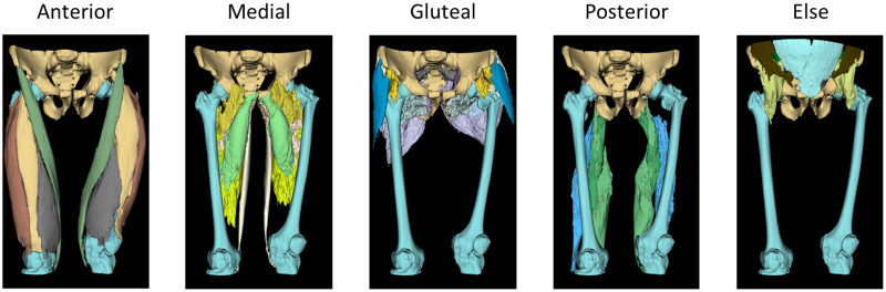
Hip fractures are a common and debilitating condition, particularly among older adults. Loss of muscle mass and strength is a common consequence of hip fractures, which further contribute to functional decline and increased disability. Assessing changes in individual thigh muscles volume in follow-up patients can provide valuable insights into the quantitative recovery process and guide rehabilitation interventions. However, accurately measuring anatomical individual thigh muscle volume can be challenging due to various, labor intensive and time-consuming.
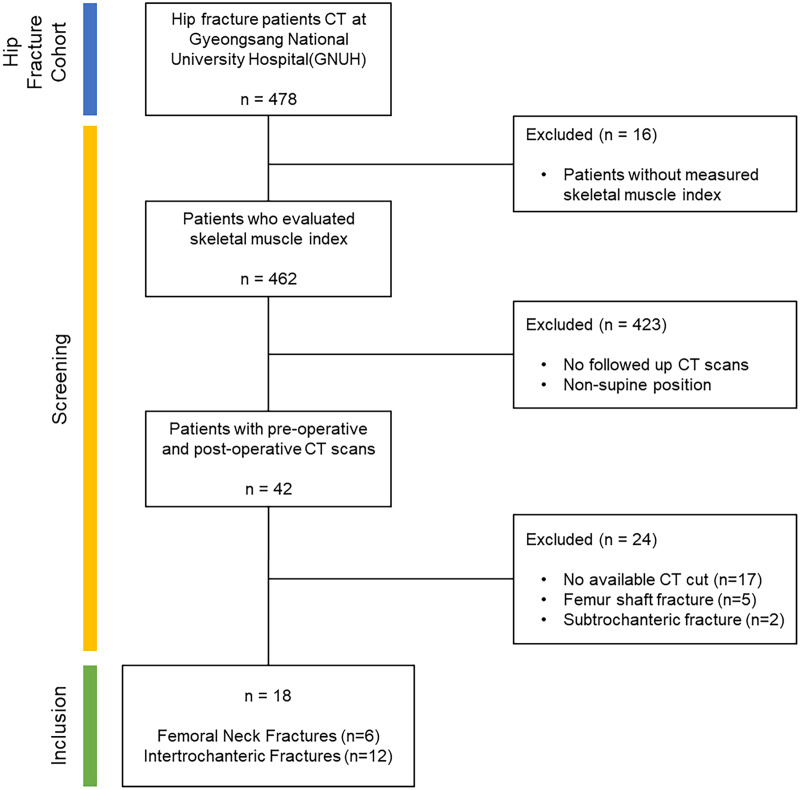
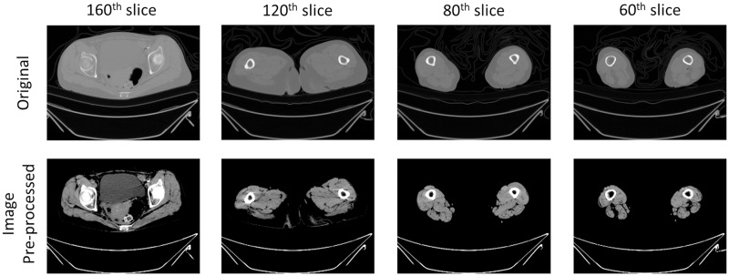
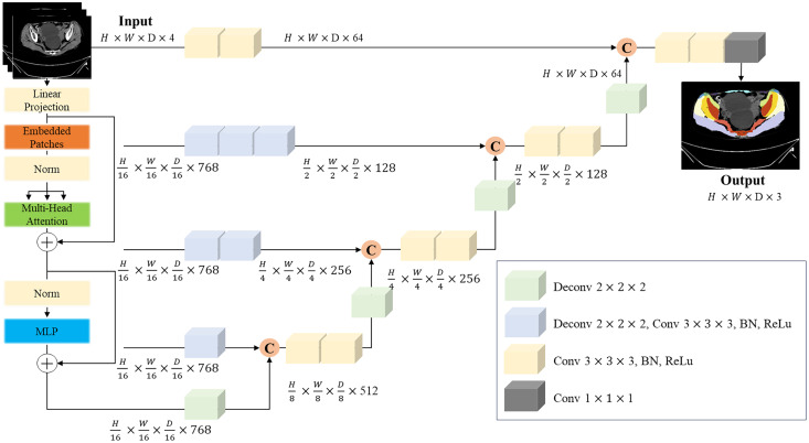
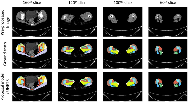
This study aimed to evaluate differences in thigh muscle volume in followed-up hip fracture patients computed tomography (CT) scans using an AI based automatic muscle segmentation model. The study included a total of 18 patients at Gyeongsang National University, who had undergone surgical treatment for a hip fracture. We utilized the automatic segmentation algorithm which we have already developed using UNETR (U-net Transformer) architecture, performance dice score = 0.84, relative absolute volume difference 0.019 ± 0.017%.
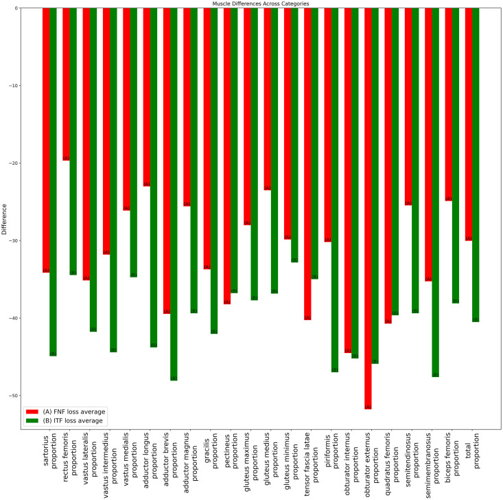
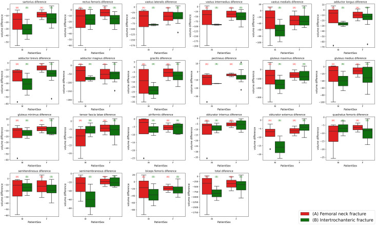
The results revealed intertrochanteric fractures result in more significant muscle volume loss (females: -97.4 cm, males: -178.2 cm) compared to femoral neck fractures (females: -83 cm, males: -147.2 cm). Additionally, the study uncovered substantial disparities in the susceptibility to volume loss among specific thigh muscles, including the Vastus lateralis, Adductor longus and brevis, and Gluteus maximus, particularly in cases of intertrochanteric fractures.
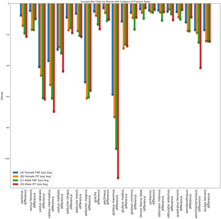
The use of an automatic muscle segmentation model based on deep learning algorithms enables efficient and accurate analysis of thigh muscle volume differences in followed up hip fracture patients. Our findings emphasize the significant muscle loss tied to sarcopenia, a critical condition among the elderly. Intertrochanteric fractures resulted in greater muscle volume deformities, especially in key muscle groups, across both genders. Notably, while most muscles exhibited volume reduction following hip fractures, the sartorius, vastus and gluteus groups demonstrated more significant disparities in individuals who sustained intertrochanteric fractures. This non-invasive approach provides valuable insights into the extent of muscle atrophy following hip fracture and can inform targeted rehabilitation interventions.
髋部骨折是一种常见且使人虚弱的疾病,特别是在老年人中。肌肉质量和力量的丧失是髋部骨折的常见后果,这进一步导致功能下降和残疾增加。在随访的髋部骨折患者中,评估个体大腿肌肉体积的变化可以提供对定量恢复过程的有价值的见解,并指导康复干预措施。然而,由于各种原因,准确测量解剖学上的个体大腿肌肉体积可能具有挑战性,例如繁琐、耗时且费力。
本研究旨在使用基于人工智能的自动肌肉分割模型评估在接受髋关节骨折随访的患者的 CT 扫描中大腿肌肉体积的差异。该研究共纳入了在全南国立大学接受髋关节骨折手术治疗的 18 名患者。我们利用了我们已经使用 UNETR(U-net Transformer)架构开发的自动分割算法,其性能骰子分数为 0.84,相对绝对体积差异为 0.019 ± 0.017%。
结果显示,与股骨颈骨折相比,转子间骨折导致更大的肌肉体积损失(女性:-97.4cm,男性:-178.2cm)(女性:-83cm,男性:-147.2cm)。此外,研究还揭示了在特定大腿肌肉中,包括股外侧肌、长内收肌和短内收肌以及臀大肌,体积损失易感性方面存在显著差异,特别是在转子间骨折的情况下。
使用基于深度学习算法的自动肌肉分割模型可以实现对髋部骨折随访患者大腿肌肉体积差异的高效准确分析。我们的发现强调了与老年人密切相关的肌肉减少症的显著肌肉损失。转子间骨折导致更大的肌肉体积畸形,特别是在两性中关键的肌肉群。值得注意的是,虽然大多数肌肉在髋部骨折后表现出体积减少,但股直肌、股外侧肌和臀大肌组在发生转子间骨折的个体中表现出更大的体积差异。这种非侵入性方法为髋部骨折后肌肉萎缩的程度提供了有价值的见解,并为有针对性的康复干预措施提供了信息。