• 文献检索
  • 文档翻译
  • 深度研究
  • 学术资讯
  • Suppr Zotero 插件Zotero 插件
  • 邀请有礼
  • 套餐&价格
  • 历史记录
应用&插件
Suppr Zotero 插件Zotero 插件浏览器插件Mac 客户端Windows 客户端微信小程序
定价
高级版会员购买积分包购买API积分包
服务
文献检索文档翻译深度研究API 文档MCP 服务
关于我们
关于 Suppr公司介绍联系我们用户协议隐私条款
关注我们

Suppr 超能文献

核心技术专利:CN118964589B侵权必究
粤ICP备2023148730 号-1Suppr @ 2026

文献检索

告别复杂PubMed语法,用中文像聊天一样搜索,搜遍4000万医学文献。AI智能推荐,让科研检索更轻松。

立即免费搜索

文件翻译

保留排版,准确专业,支持PDF/Word/PPT等文件格式,支持 12+语言互译。

免费翻译文档

深度研究

AI帮你快速写综述,25分钟生成高质量综述,智能提取关键信息,辅助科研写作。

立即免费体验

上颌和下颌前磨牙氧化锆全瓷内冠的颜色变化、边缘适合性、断裂强度和失效类型的评价。

Evaluation of colour change, marginal adaptation, fracture strength, and failure type in maxillary and mandibular premolar zirconia endo-crowns.

机构信息

Department of Restorative Dental Science, College of Dentistry, Jazan University, Jazan, 45142, Saudi Arabia.

Department of Prosthetic Dental Science, College of Dentistry, Jazan University, Jazan, 45142, Saudi Arabia.

出版信息

BMC Oral Health. 2024 Aug 21;24(1):970. doi: 10.1186/s12903-024-04755-z.

DOI:10.1186/s12903-024-04755-z
PMID:39169344
原文链接:https://pmc.ncbi.nlm.nih.gov/articles/PMC11340096/
Abstract

OBJECTIVES

The objective of this in vitro study was to evaluate the effects of different preparation designs on the mean colour change (ΔE), marginal adaptation, fracture resistance, and fracture types of maxillary and mandibular premolar endocrowns (ECs).

METHODOLOGY

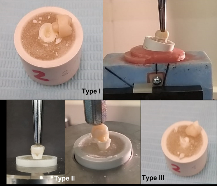
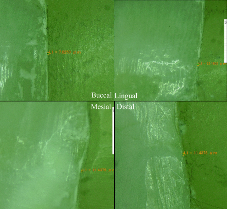
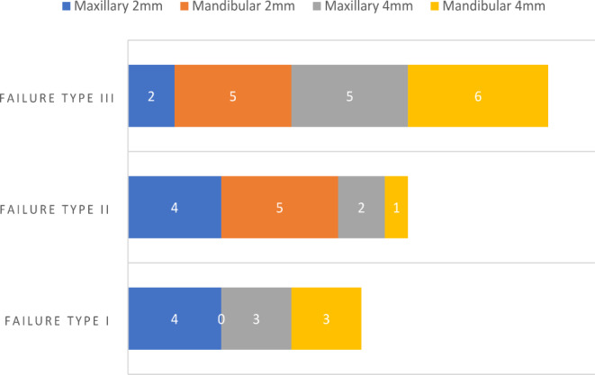
A total of 40 extracted maxillary and mandibular premolars were treated endodontically, and each type was subdivided according to the remaining axial height (remaining walls on all surfaces; 2-4 mm) and 2 mm inside the pulp chamber. Specimens were immersed in coffee for 14 days, ΔE was determined, marginal adaptation was observed, fracture forces test was conducted, and the samples were examined visually at 10× magnification to evaluate failure type and identify fracture origin. The data were entered and analyzed using Statistical Package for Social Sciences, and significance between and within groups was evaluated through ANOVA. The p-value ≤ 0.05 was considered statistically significant.

RESULTS

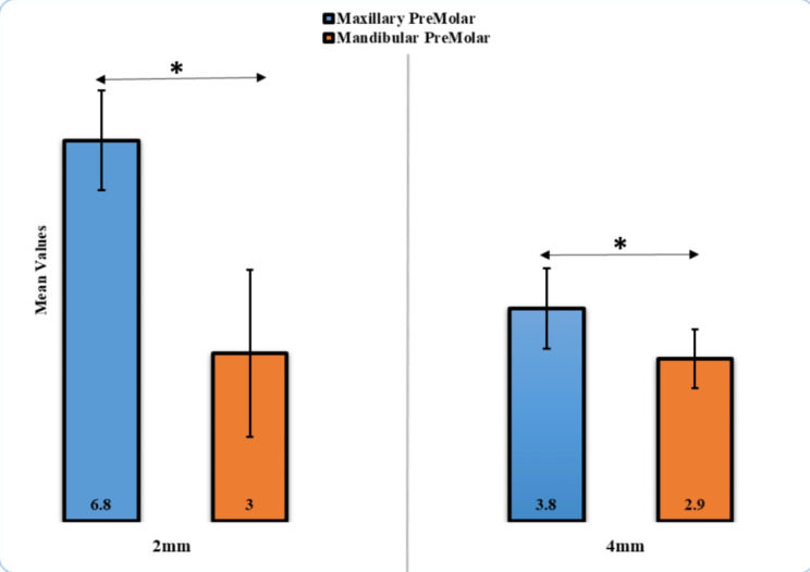
The ΔE* values of the maxillary premolar with 2 mm axial height were the highest (6.8 ± 0.89 units), whereas the lowest value was observed in the mandibular premolar with 4 mm axial height (2.9 ± 0.53 units). Significant differences (p < 0.05) in teeth and design were observed. The marginal adaptation of the mandibular premolar with 4 mm axial height was the highest (30.20 ± 1.53 μm), whereas the lowest marginal adaptation was observed in the maxillary premolar with 2 mm axial height (14.38 ± 0.99 μm), and the difference was statistically significant (p < 0.05). The maximum fracture force was observed in maxillary premolars with 2 mm axial height (2248.15 ± 134.74 N), and no statistically significant difference (p = 0.07) was observed between maxillary and mandibular premolars at 4 mm axial height.

CONCLUSION

The recorded ΔE values of the ECs were within clinically acceptable values or slightly higher, and the marginal adaption values were within acceptable and recommended clinical values in µm. EC preparation with 2 mm axial height in both arches recorded the highest fracture forces. Type III (split fracture) failure was recorded as the highest in the maxillary and mandibular premolar ECs with different axial wall heights.

摘要

目的

本体外研究旨在评估不同预备设计对上颌和下颌前磨牙嵌体冠(EC)的平均颜色变化(ΔE)、边缘适合性、抗折强度和骨折类型的影响。

方法

共处理 40 颗上颌和下颌前磨牙的牙髓,并根据剩余轴向高度(所有表面的剩余壁;2-4mm)和牙髓腔内部的 2mm 将每种类型进一步细分。将标本浸入咖啡中 14 天,测定 ΔE,观察边缘适合性,进行抗折强度测试,并在 10×放大倍数下目视检查样品,以评估失效类型并确定骨折起源。使用社会科学统计软件包输入和分析数据,并通过方差分析评估组间和组内的显著性。p 值≤0.05 被认为具有统计学意义。

结果

2mm 轴向高度的上颌前磨牙的 ΔE*值最高(6.8±0.89 单位),而 4mm 轴向高度的下颌前磨牙的最低值(2.9±0.53 单位)。牙齿和设计之间存在显著差异(p<0.05)。4mm 轴向高度的下颌前磨牙的边缘适合性最高(30.20±1.53μm),而 2mm 轴向高度的上颌前磨牙的最低边缘适合性(14.38±0.99μm),差异具有统计学意义(p<0.05)。2mm 轴向高度的上颌前磨牙的最大骨折力最高(2248.15±134.74N),4mm 轴向高度的上颌和下颌前磨牙之间无统计学显著差异(p=0.07)。

结论

EC 的记录 ΔE 值在临床可接受范围内或略高,边缘适应值在 µm 内处于可接受和推荐的临床值范围内。上颌和下颌的 2mm 轴向高度的 EC 预备记录了最高的骨折力。在上颌和下颌前磨牙 EC 中,记录到的 III 型(劈裂骨折)失效最高。

https://cdn.ncbi.nlm.nih.gov/pmc/blobs/9c7f/11340096/a68ada3ea94e/12903_2024_4755_Fig8_HTML.jpg
https://cdn.ncbi.nlm.nih.gov/pmc/blobs/9c7f/11340096/0b5cb72ddeb2/12903_2024_4755_Fig1_HTML.jpg
https://cdn.ncbi.nlm.nih.gov/pmc/blobs/9c7f/11340096/681e7f0d21ff/12903_2024_4755_Fig2_HTML.jpg
https://cdn.ncbi.nlm.nih.gov/pmc/blobs/9c7f/11340096/5cdc1cf0251c/12903_2024_4755_Fig3_HTML.jpg
https://cdn.ncbi.nlm.nih.gov/pmc/blobs/9c7f/11340096/59ef5c8b15a0/12903_2024_4755_Fig4_HTML.jpg
https://cdn.ncbi.nlm.nih.gov/pmc/blobs/9c7f/11340096/7c43861af925/12903_2024_4755_Fig5_HTML.jpg
https://cdn.ncbi.nlm.nih.gov/pmc/blobs/9c7f/11340096/95bc18470256/12903_2024_4755_Fig6_HTML.jpg
https://cdn.ncbi.nlm.nih.gov/pmc/blobs/9c7f/11340096/585fcac763dc/12903_2024_4755_Fig7_HTML.jpg
https://cdn.ncbi.nlm.nih.gov/pmc/blobs/9c7f/11340096/a68ada3ea94e/12903_2024_4755_Fig8_HTML.jpg

相似文献

1
Evaluation of colour change, marginal adaptation, fracture strength, and failure type in maxillary and mandibular premolar zirconia endo-crowns.上颌和下颌前磨牙氧化锆全瓷内冠的颜色变化、边缘适合性、断裂强度和失效类型的评价。
BMC Oral Health. 2024 Aug 21;24(1):970. doi: 10.1186/s12903-024-04755-z.
2
Comparison of color changes, fracture strengths, and failure modes of conventional endocrowns and endocrowns with different design modifications.传统嵌体冠与不同设计改良嵌体冠的颜色变化、断裂强度及失效模式的比较。
Technol Health Care. 2024;32(4):2395-2408. doi: 10.3233/THC-231175.
3
Marginal adaptation and fracture resistance of feldspathic and polymer-infiltrated ceramic network CAD/CAM endocrowns for maxillary premolars.用于上颌前磨牙的长石质和聚合物渗透陶瓷网络CAD/CAM内冠的边缘适应性和抗折性
Niger J Clin Pract. 2020 Jan;23(1):1-6. doi: 10.4103/njcp.njcp_231_19.
4
Influence of the type of post and core on in vitro marginal continuity, fracture resistance, and fracture mode of lithia disilicate-based all-ceramic crowns.桩核类型对硅酸锂基全瓷冠体外边缘密合性、抗折性及折裂模式的影响。
J Prosthet Dent. 2008 Oct;100(4):264-73. doi: 10.1016/S0022-3913(08)60205-X.
5
Effect of fiber post length and abutment height on fracture resistance of endodontically treated premolars prepared for zirconia crowns.纤维桩长度和基牙高度对为氧化锆全冠修复而进行根管治疗的前磨牙抗折性能的影响。
Odontology. 2018 Apr;106(2):215-222. doi: 10.1007/s10266-017-0320-7. Epub 2017 Dec 14.
6
Rehabilitation of severely-destructed endodontically treated premolar teeth with novel endocrown system: Biomechanical behavior assessment through 3D finite element and in vitro analyses.使用新型根管内冠系统修复严重破坏的根管治疗前磨牙:通过三维有限元分析和体外分析进行生物力学行为评估
J Mech Behav Biomed Mater. 2022 Feb;126:105031. doi: 10.1016/j.jmbbm.2021.105031. Epub 2021 Dec 10.
7
Fracture strength of endocrown maxillary restorations using different preparation designs and materials.上颌牙内冠修复不同预备设计和材料的断裂强度。
J Mech Behav Biomed Mater. 2023 Dec;148:106184. doi: 10.1016/j.jmbbm.2023.106184. Epub 2023 Oct 10.
8
Comparative evaluation of marginal adaptation and fatigue resistance of endodontically treated premolars restored with direct and indirect coronal restorations: an in vitro study.根管治疗后前磨牙的直接和间接冠部修复体的边缘适合性和耐疲劳性的比较评价:一项体外研究。
BMC Oral Health. 2024 Jun 15;24(1):696. doi: 10.1186/s12903-024-04382-8.
9
Fracture resistance and failure modes of polymer infiltrated ceramic endocrown restorations with variations in margin design and occlusal thickness.聚合物渗透陶瓷嵌体修复体的断裂阻力和失效模式,其边缘设计和咬合厚度存在差异。
J Prosthodont Res. 2018 Jul;62(3):293-297. doi: 10.1016/j.jpor.2017.11.003. Epub 2017 Dec 11.
10
The influence of zirconia and fiber posts with different lengths on the fracture strength of maxillary central incisors restored with zirconia crowns: an in vitro study.氧化锆和不同长度纤维桩对上颌中切牙全瓷冠修复后抗折强度的影响:一项体外研究。
Int J Comput Dent. 2024 Jun 6;27(2):161-167. doi: 10.3290/j.ijcd.b3963385.

引用本文的文献

1
Experimental study on the compressive strength of fixed prosthetic restorations on tooth preparations with or without horizontal preparation.有或无水平预备的牙体预备上固定修复体抗压强度的实验研究
J Med Life. 2025 Jul;18(7):671-677. doi: 10.25122/jml-2025-0124.
2
Biomechanical and Occlusal Factors Influencing the Longevity of Single-Unit Restorations: A Comprehensive Review.影响单单位修复体使用寿命的生物力学和咬合因素:一项综述。
Cureus. 2025 Jun 14;17(6):e85998. doi: 10.7759/cureus.85998. eCollection 2025 Jun.
3
Effect of Pulpal Base, Restorative Material, and Preparation Type on Marginal and Internal Fit and Fracture Strength of Endocrowns.

本文引用的文献

1
Comparison of color changes, fracture strengths, and failure modes of conventional endocrowns and endocrowns with different design modifications.传统嵌体冠与不同设计改良嵌体冠的颜色变化、断裂强度及失效模式的比较。
Technol Health Care. 2024;32(4):2395-2408. doi: 10.3233/THC-231175.
2
A Comparative Analysis of Marginal Adaptation Values between Lithium Disilicate Glass Ceramics and Zirconia-Reinforced Lithium Silicate Endocrowns: A Systematic Review of In Vitro Studies.硅酸镧锂玻璃陶瓷与氧化锆增强硅酸锂全瓷冠边缘适合性的比较分析:体外研究的系统评价
Med Sci Monit. 2023 Dec 27;29:e942649. doi: 10.12659/MSM.942649.
3
牙髓基底、修复材料及预备类型对内冠边缘与内部适合性及抗折强度的影响
Materials (Basel). 2025 May 6;18(9):2137. doi: 10.3390/ma18092137.
4
Impact of Thermal Aging on Marginal Adaptation in Lithium Disilicate CAD/CAM Crowns with Deep Proximal Box Elevation.热老化对具有深邻面盒提升的二硅酸锂CAD/CAM全冠边缘适合性的影响。
Med Sci Monit. 2025 Feb 3;31:e947191. doi: 10.12659/MSM.947191.
Fracture strength of endocrown maxillary restorations using different preparation designs and materials.
上颌牙内冠修复不同预备设计和材料的断裂强度。
J Mech Behav Biomed Mater. 2023 Dec;148:106184. doi: 10.1016/j.jmbbm.2023.106184. Epub 2023 Oct 10.
4
Relationship between tooth type and material used in the construction of endocrowns and fracture force values: a systematic review.牙体类型与核冠修复材料和抗折力值的关系:系统评价。
Eur Rev Med Pharmacol Sci. 2023 Aug;27(16):7665-7679. doi: 10.26355/eurrev_202308_33420.
5
Unlocking Endocrown Restoration Expertise Among Dentists: Insights from a Multi-Center Cross-Sectional Study.解锁牙医的后牙全瓷嵌体修复专长:一项多中心横断面研究的启示。
Med Sci Monit. 2023 Jun 29;29:e940573. doi: 10.12659/MSM.940573.
6
Biomechanical behavior of all-ceramic endocrowns fabricated using CAD/CAM: A systematic review.CAD/CAM 制作的全瓷内冠的生物力学行为:系统评价。
J Prosthodont Res. 2024 Jan 16;68(1):50-62. doi: 10.2186/jpr.JPR_D_22_00296. Epub 2023 Jun 7.
7
In-Vitro Investigation of Marginal Adaptation and Fracture Resistance of Resin Matrix Ceramic Endo-Crown Restorations.树脂基陶瓷根管内冠修复体边缘适应性和抗折性的体外研究
Materials (Basel). 2023 Mar 2;16(5):2059. doi: 10.3390/ma16052059.
8
Endocrown restorations in premolars: influence of remaining axial walls of tooth structure and restorative materials on fatigue resistance.后磨牙的嵌体修复:牙体结构剩余轴壁和修复材料对疲劳强度的影响。
Clin Oral Investig. 2023 Jun;27(6):2957-2968. doi: 10.1007/s00784-023-04895-6. Epub 2023 Feb 14.
9
Fracture resistance of resin endocrowns with and without fiber reinforced composite base material: A preliminary study.含与不含纤维增强复合材料基底材料的树脂桩核冠的抗折性:一项初步研究。
J Appl Biomater Funct Mater. 2023 Jan-Dec;21:22808000231151832. doi: 10.1177/22808000231151832.
10
Effect of thickness on the translucency of resin-based composites and glass-ceramics.厚度对树脂基复合材料和玻璃陶瓷透明度的影响。
Dent Mater J. 2023 Jan 31;42(1):30-41. doi: 10.4012/dmj.2022-093. Epub 2022 Oct 14.