Veterinary Medicine, Universidade de Franca (UNIFRAN), Franca, São Paulo, Brazil.
Veterinary Surgery, Veterinary Hospital, Universidade de Franca (UNIFRAN), Franca, São Paulo, Brazil.
Open Vet J. 2024 Jul;14(7):1708-1715. doi: 10.5455/OVJ.2024.v14.i7.20. Epub 2024 Jul 31.
Primary ureteral neoplasms are extremely rare in dogs, and ureteral involvement usually occurs owing to the invasion of renal and bladder tumors.
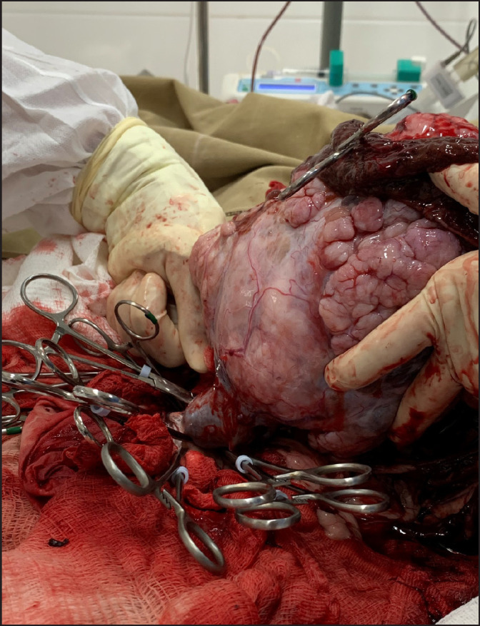
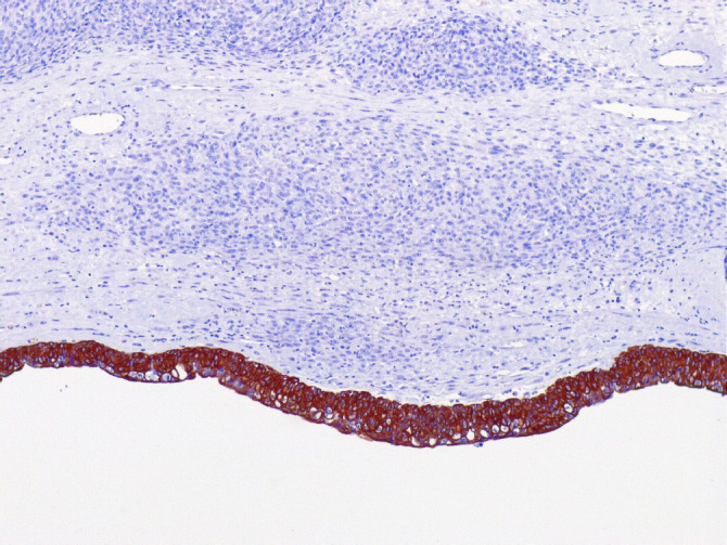
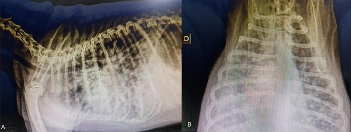
This case report describes a 12-year-old intact male mixed-breed dog referred to a private clinic with a six-month history of abdominal distention. A physical examination revealed mild abdominal pain. Hematological tests detected normocytic-normochromic anemia (hematocrit 33.6% [reference interval-RI: 37%-55%], red blood cells 4.93 M/µl [RI: 5.5-8.5 M/µl], and hemoglobin 12.4 g/dl [RI: 12-18.0 g/dl]). The results from the leukogram, thrombogram, renal, and hepatic panels were within the reference intervals for dogs. Abdominal ultrasonography revealed a cavitary mass measuring approximately 12 cm in diameter as the largest tumor in the left abdominal region over the left hepatic lobe or mesenteric site. Chest radiography did not reveal any metastasis. Therefore, the patient underwent exploratory laparotomy, during which the left ureter was found to be affected by a 12-cm mass that adhered to the left kidney. A unilateral left ureteronephrectomy was performed, and histology and immunohistochemistry (IHC) confirmed well-differentiated primary ureteral leiomyosarcoma. The patient survived for 130 days but died of lung metastasis.
Ureteral leiomyosarcoma should be investigated and included in the list of differential diagnoses for primary ureteral neoplasms. Regardless of the therapeutic modality, the prognosis of ureteral leiomyosarcoma may be unfavorable, as shown in this report.
原发性输尿管肿瘤在犬中极为罕见,输尿管受累通常是由于肾和膀胱肿瘤的侵袭所致。
本病例报告描述了一例 12 岁未绝育雄性混血犬,因腹部膨胀就诊于一家私人诊所,病史为 6 个月。体格检查发现轻度腹痛。血液学检查发现正细胞正色素性贫血(血细胞比容 33.6%[参考区间-RI:37%-55%],红细胞 4.93×10^6/µl[RI:5.5-8.5×10^6/µl],血红蛋白 12.4g/dl[RI:12-18.0g/dl])。白细胞计数、血小板计数、肾功能和肝功能检查结果均在犬的参考区间内。腹部超声检查显示左肝叶或肠系膜部位左腹部有一个最大直径约 12cm 的腔隙性肿块,为最大肿瘤。胸部 X 线摄影未发现任何转移。因此,患者接受了剖腹探查术,术中发现左输尿管受一个 12cm 大的肿块影响,该肿块与左肾粘连。进行了单侧左输尿管肾切除术,组织学和免疫组织化学(IHC)证实为分化良好的原发性输尿管平滑肌肉瘤。该患者存活 130 天,但死于肺转移。
应调查并将输尿管平滑肌肉瘤纳入原发性输尿管肿瘤的鉴别诊断清单中。无论采用何种治疗方式,输尿管平滑肌肉瘤的预后可能都不理想,正如本报告所示。