Department of Chemistry, Texas A&M University, College Station, Texas, USA.
Department of Biochemistry and Biophysics, Texas A&M University, College Station, Texas, USA.
J Biol Chem. 2024 Sep;300(9):107711. doi: 10.1016/j.jbc.2024.107711. Epub 2024 Aug 22.
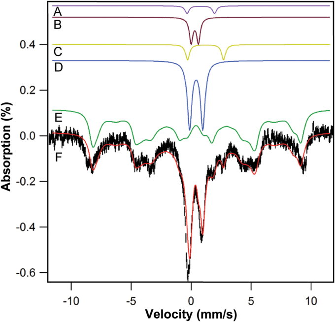
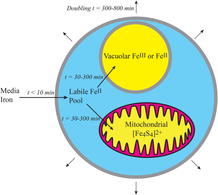
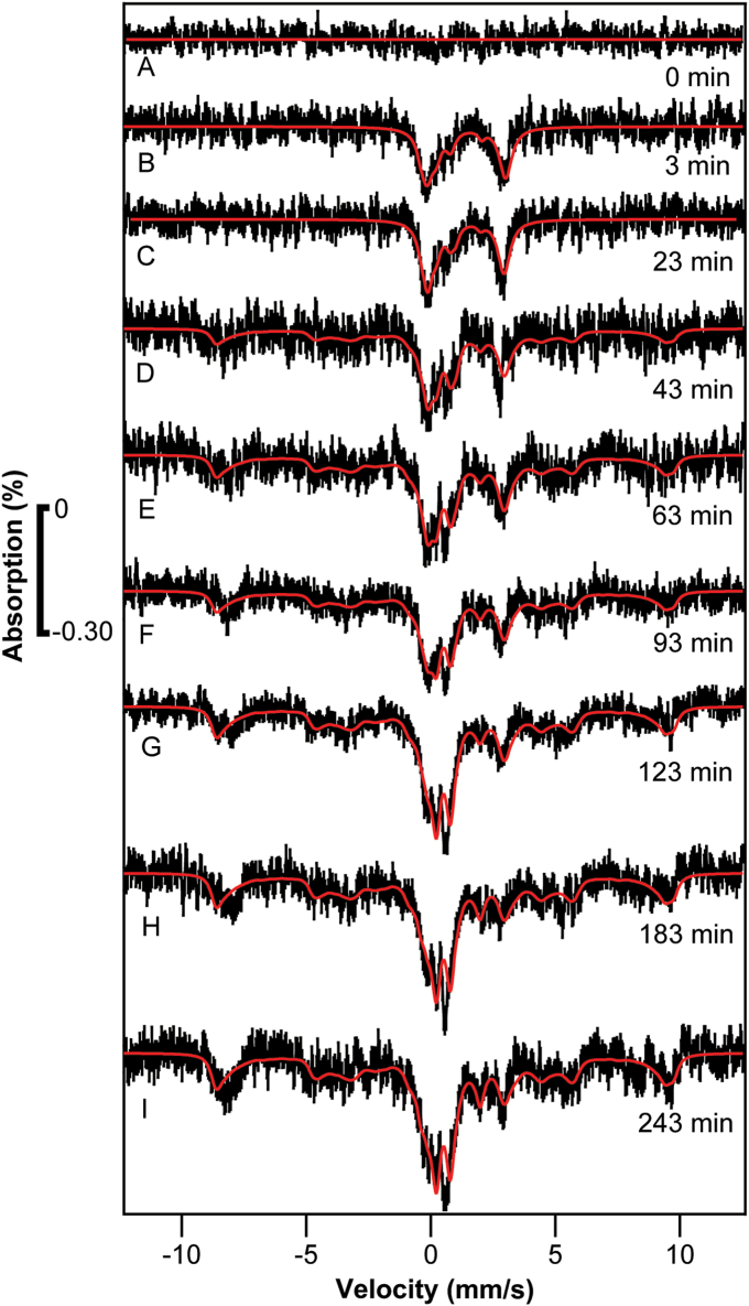
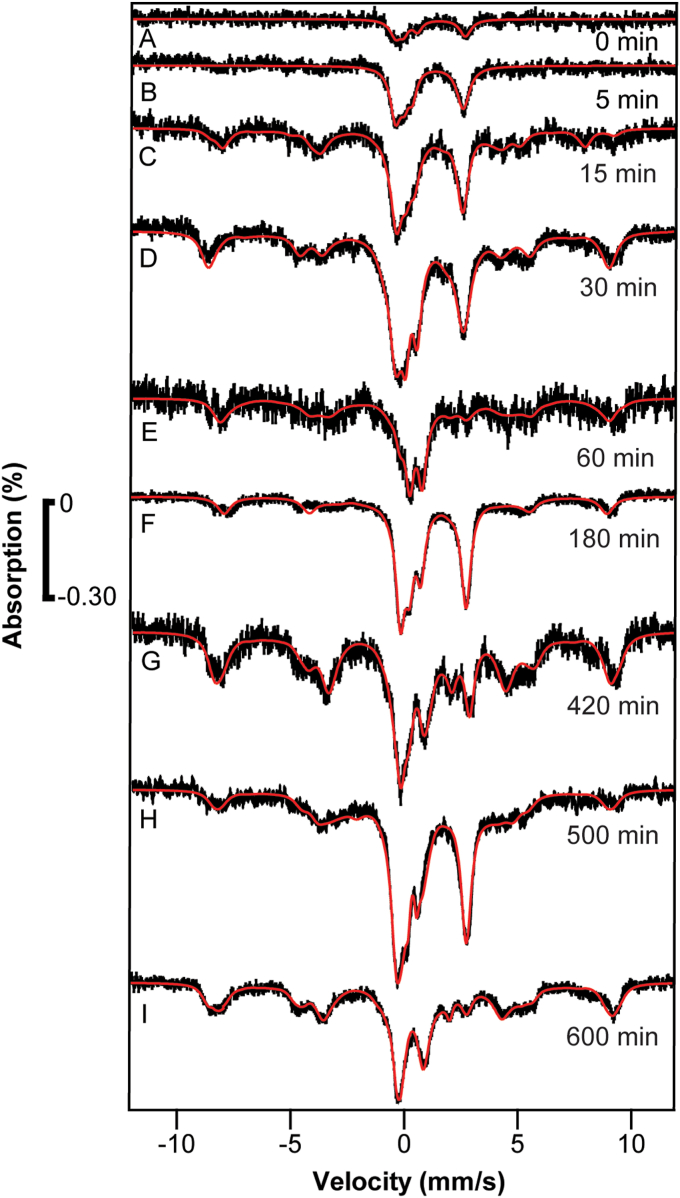
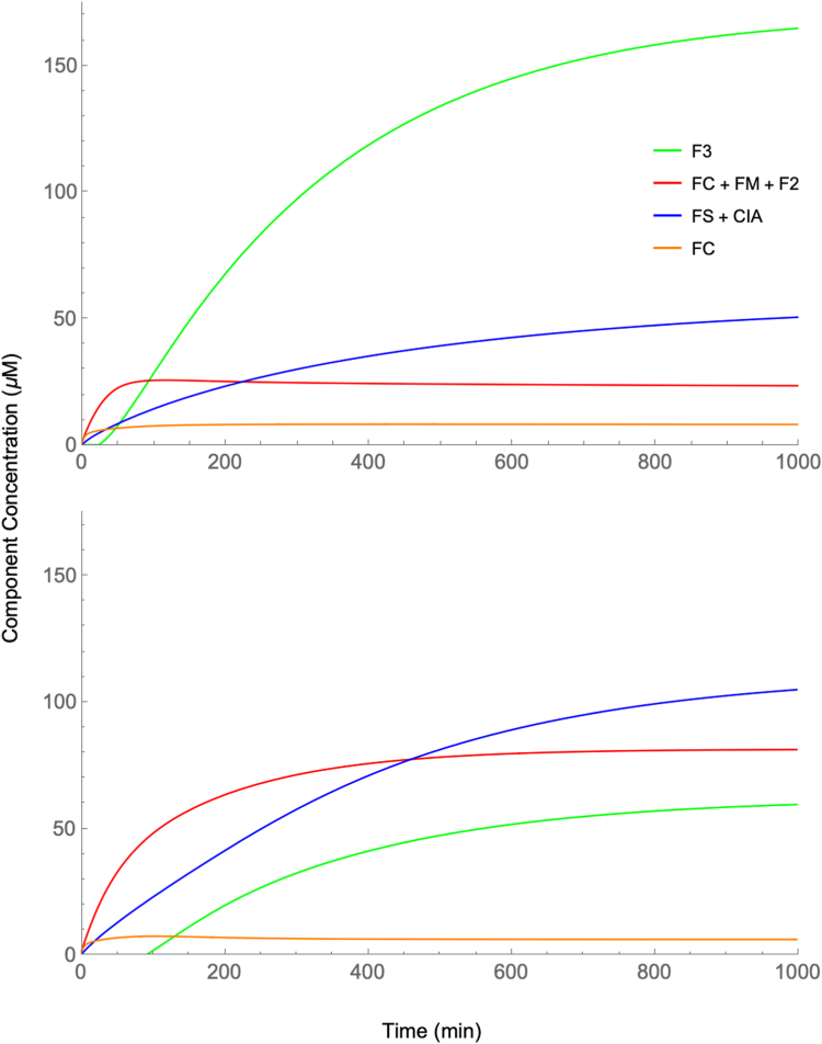
The kinetics of iron trafficking in whole respiring Saccharomyces cerevisiae cells were investigated using Mössbauer and EPR spectroscopies. The Mössbauer-active isotope Fe was added to cells growing under iron-limited conditions; cells were analyzed at different times post iron addition. Spectroscopic changes suggested that the added Fe initially entered the labile iron pool, and then distributed to vacuoles and mitochondria. The first spectroscopic feature observed, ∼ 3 min after adding Fe plus a 5 to 15 min processing dead time, was a quadrupole doublet typical of nonheme high-spin Fe. This feature likely arose from labile Fe pools in the cell. At later times (15-150 min), magnetic features due to S = 5/2 Fe developed; these likely arose from Fe in vacuoles. Corresponding EPR spectra were dominated by a g = 4.3 signal from the S = 5/2 Fe ions that increased in intensity over time. Developing at a similar rate was a quadrupole doublet typical of S = 0 [FeS] clusters and low-spin Fe hemes; such centers are mainly in mitochondria, cytosol, and nuclei. Development of these features was simulated using a published mathematical model, and simulations compared qualitatively well with observations. In the five sets of experiments presented, all spectroscopic features developed within the doubling time of the cells, implying that the detected iron trafficking species are physiologically relevant. These spectroscopy-based experiments allow the endogenous labile iron pool within growing cells to be detected without damaging or altering the pool, as definitely occurs using chelator-probe detection and possibly occurs using chromatographic separations.
使用 Mössbauer 和 EPR 光谱研究了整个呼吸酿酒酵母细胞中铁转运的动力学。将 Mössbauer 活性同位素 Fe 添加到铁限制条件下生长的细胞中;在添加铁后不同时间分析细胞。光谱变化表明,添加的 Fe 最初进入不稳定铁池,然后分布到液泡和线粒体。添加 Fe 后观察到的第一个光谱特征是在 3 分钟左右,存在典型的非血红素高自旋 Fe 的四极子双峰。此特征可能来自细胞中的不稳定铁池。在稍后的时间(15-150 分钟),由于 S = 5/2 Fe 而出现磁性特征;这些可能来自液泡中的 Fe。相应的 EPR 光谱主要由 g = 4.3 信号主导,该信号来自 S = 5/2 Fe 离子,随着时间的推移其强度增加。以相似的速率发展的是典型的 S = 0 [FeS]簇和低自旋 Fe 血红素的四极子双峰;这些中心主要存在于线粒体、细胞质和核中。使用已发表的数学模型模拟了这些特征的发展,模拟与观察结果定性上吻合较好。在所呈现的五组实验中,所有光谱特征都在细胞倍增时间内发展,这意味着检测到的铁转运物质在生理上是相关的。这些基于光谱的实验允许在不破坏或改变池的情况下检测到生长细胞内的内源性不稳定铁池,这肯定会发生在使用螯合剂探针检测时,也可能会发生在使用色谱分离时。