• 文献检索
  • 文档翻译
  • 深度研究
  • 学术资讯
  • Suppr Zotero 插件Zotero 插件
  • 邀请有礼
  • 套餐&价格
  • 历史记录
应用&插件
Suppr Zotero 插件Zotero 插件浏览器插件Mac 客户端Windows 客户端微信小程序
定价
高级版会员购买积分包购买API积分包
服务
文献检索文档翻译深度研究API 文档MCP 服务
关于我们
关于 Suppr公司介绍联系我们用户协议隐私条款
关注我们

Suppr 超能文献

核心技术专利:CN118964589B侵权必究
粤ICP备2023148730 号-1Suppr @ 2026

文献检索

告别复杂PubMed语法,用中文像聊天一样搜索,搜遍4000万医学文献。AI智能推荐,让科研检索更轻松。

立即免费搜索

文件翻译

保留排版,准确专业,支持PDF/Word/PPT等文件格式,支持 12+语言互译。

免费翻译文档

深度研究

AI帮你快速写综述,25分钟生成高质量综述,智能提取关键信息,辅助科研写作。

立即免费体验

采用跗骨中部截骨联合伊里扎洛夫技术矫正僵硬性马蹄高弓足

The Gradual Correction of Rigid Pes Cavus Using Midfoot Osteotomy Combined with Ilizarov Methods.

机构信息

Department of Orthopedics, Orthopedic Research Institute, West China Hospital, Sichuan University, Chengdu, China.

出版信息

Orthop Surg. 2024 Sep;16(9):2242-2251. doi: 10.1111/os.14206. Epub 2024 Aug 26.

DOI:10.1111/os.14206
PMID:39187990
原文链接:https://pmc.ncbi.nlm.nih.gov/articles/PMC11572576/
Abstract

OBJECTIVE

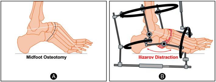
Midfoot osteotomy combined with Ilizarov methods of correction is a rarely reported treatment that is particularly well-suited for severe rigid pes cavus. The study aimed to assess the radiological and clinical results of patients who had been treated for rigid pes cavus using this method.

METHODS

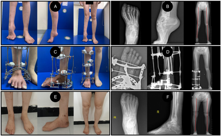
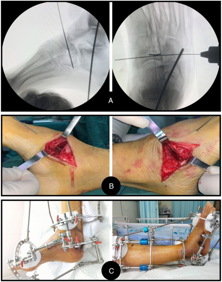
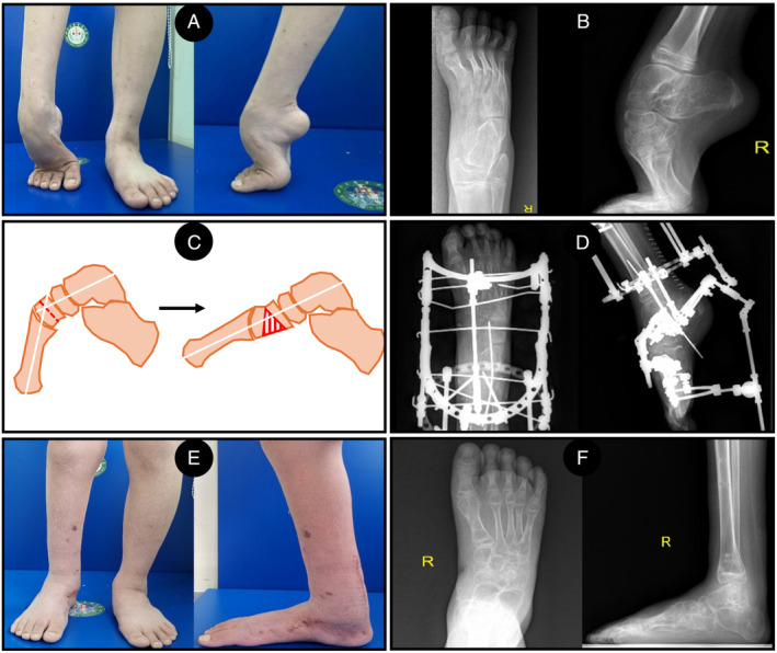
The study retrospectively analyzed the clinical and radiological data of 15 pes cavus in 12 patients who were corrected by midfoot osteotomy with Ilizarov external frame in our department from March 2020 to September 2022. Radiologic outcomes were measured using the Meary angle (MA), talus-first metatarsal angle (TM1A), calcaneal varus angle (CVA) and foot length with weight-bearing radiographs. Functional assessments were evaluated in terms of pain, function, and quality of life by using the visual analogue scale (VAS), the American Orthopedic Foot and Ankle Society hindfoot scale score (AOFAS), and 36-item Short Form Health Survey (SF-36). Additionally, the postoperative satisfaction of patients was investigated by a questionnaire. The clinical and radiological results were evaluated by a paired t-test.

RESULTS

All patients received plantigrade feet and pain relief. The mean follow-up was 33.1 ± 5.0 months (range from 25 to 41 months). The etiology included poliomyelitis (4), idiopathic (3), trauma (2), spina bifida (2) and tethered cord syndrome (1). The duration of gradual correction was 30.4 ± 10.6 days, and the external fixation time was 116.3 ± 33.3 days. The bony union rate was 100%. The VAS, AOFAS, and SF-36 scores significantly improved (p < 0.05). The MA, TM1A, and CVA were close to or reached the normal range postoperative (p < 0.01). The length of each foot was well preserved, which was increased more than 0.8 cm than preoperative. No major complications were reported except two cases of mildly hindfoot varus deformity. The results of the questionnaire showed that patients' satisfaction was 92% (11/12).

CONCLUSION

Midfoot osteotomy combined with Ilizarov external frame proved to be a reasonable procedure with satisfying mid-term results for the gradual correction of rigid pes cavus.

摘要

目的

中足截骨联合伊利扎洛夫矫正方法是一种很少报道的治疗方法,特别适合严重僵硬性马蹄内翻足。本研究旨在评估采用该方法治疗僵硬性马蹄内翻足患者的放射学和临床结果。

方法

本研究回顾性分析了 2020 年 3 月至 2022 年 9 月我科采用中足截骨联合伊利扎洛夫外固定架治疗的 12 例 15 例僵硬性马蹄内翻足患者的临床和放射学资料。采用 Meary 角(MA)、距骨第一跖骨角(TM1A)、跟骨内翻角(CVA)和负重位足部长度测量放射学结果。采用视觉模拟评分(VAS)、美国矫形足踝协会后足评分(AOFAS)和 36 项简明健康状况调查问卷(SF-36)评估疼痛、功能和生活质量。此外,通过问卷调查调查了患者的术后满意度。采用配对 t 检验评估临床和放射学结果。

结果

所有患者均获得足底负重和缓解疼痛。平均随访 33.1±5.0 个月(25~41 个月)。病因包括脊髓灰质炎(4 例)、特发性(3 例)、创伤(2 例)、脊髓脊膜膨出(2 例)和脊髓栓系综合征(1 例)。逐渐矫正的时间为 30.4±10.6 天,外固定时间为 116.3±33.3 天。骨愈合率为 100%。VAS、AOFAS 和 SF-36 评分均显著改善(p<0.05)。MA、TM1A 和 CVA 术后接近或达到正常范围(p<0.01)。每只脚的长度都得到了很好的保留,比术前增加了 0.8 厘米以上。除 2 例轻度后足内翻畸形外,无其他重大并发症。问卷调查结果显示,患者满意度为 92%(11/12)。

结论

中足截骨联合伊利扎洛夫外固定架逐渐矫正僵硬性马蹄内翻足是一种合理的方法,中期结果满意。

https://cdn.ncbi.nlm.nih.gov/pmc/blobs/8a37/11572576/5e3fc9bc247b/OS-16-2242-g005.jpg
https://cdn.ncbi.nlm.nih.gov/pmc/blobs/8a37/11572576/6d0b908efee2/OS-16-2242-g001.jpg
https://cdn.ncbi.nlm.nih.gov/pmc/blobs/8a37/11572576/fbed382c37e1/OS-16-2242-g003.jpg
https://cdn.ncbi.nlm.nih.gov/pmc/blobs/8a37/11572576/a6026b9a3006/OS-16-2242-g004.jpg
https://cdn.ncbi.nlm.nih.gov/pmc/blobs/8a37/11572576/5e3fc9bc247b/OS-16-2242-g005.jpg

相似文献

1
The Gradual Correction of Rigid Pes Cavus Using Midfoot Osteotomy Combined with Ilizarov Methods.采用跗骨中部截骨联合伊里扎洛夫技术矫正僵硬性马蹄高弓足
Orthop Surg. 2024 Sep;16(9):2242-2251. doi: 10.1111/os.14206. Epub 2024 Aug 26.
2
The Cole Midfoot Osteotomy: Clinical and Radiographic Retrospective Review of Five Patients (Six Feet) with Different Etiologies.科尔中足截骨术:对五例(六足)不同病因患者的临床及影像学回顾性研究
J Am Podiatr Med Assoc. 2019 May;109(3):180-186. doi: 10.7547/17-056.
3
The Gradual Correction of Adult Severe Rigid Equinus Deformity Using Minimal Invasive U-Osteotomy With Taylor Spatial Frame.使用泰勒空间框架的微创U形截骨术逐步矫正成人重度僵硬马蹄内翻畸形
Foot Ankle Int. 2023 Jan;44(1):62-70. doi: 10.1177/10711007221138240. Epub 2022 Dec 20.
4
A prospective study of midfoot osteotomy combined with adjacent joint sparing internal fixation in treatment of rigid pes cavus deformity.一项关于中足截骨术联合保留相邻关节内固定治疗僵硬型高弓足畸形的前瞻性研究。
J Orthop Surg Res. 2014 Jun 5;9:44. doi: 10.1186/1749-799X-9-44.
5
Cavovarus foot treated with combined calcaneus and metatarsal osteotomies.采用跟骨和跖骨联合截骨术治疗高弓足。
Foot Ankle Int. 2001 Jan;22(1):19-30. doi: 10.1177/107110070102200104.
6
Flexible cavovarus feet in Charcot-Marie-Tooth disease treated with first ray proximal dorsiflexion osteotomy combined with soft tissue surgery: a short-term to mid-term outcome study.Charcot-Marie-Tooth 病中足的第一跖骨近端背屈截骨术联合软组织手术治疗:短期至中期疗效研究
Foot Ankle Surg. 2010 Sep;16(3):142-7. doi: 10.1016/j.fas.2009.10.002.
7
Results of operative correction of grade IIB tibialis posterior tendon dysfunction.IIB级胫后肌腱功能障碍的手术矫正结果。
Foot Ankle Int. 2015 Feb;36(2):165-71. doi: 10.1177/1071100714556758. Epub 2014 Nov 17.
8
Pes Cavus高弓足
9
Focal Dome Osteotomy Combined with Ilizarov Technique for Treating Femoral Multiplanar Deformity.焦点穹顶截骨术联合伊利扎洛夫技术治疗股骨多平面畸形。
Orthop Surg. 2024 Sep;16(9):2230-2241. doi: 10.1111/os.14190. Epub 2024 Aug 26.
10
Is Ankle Arthrodesis With an Ilizarov External Fixator an Effective Treatment for Septic Ankle Arthritis? A Study With a Minimum of 6 Years of Follow-up.采用伊里扎洛夫外固定架踝关节融合术治疗感染性踝关节关节炎是否有效?一项至少 6 年随访的研究。
Clin Orthop Relat Res. 2023 Apr 1;481(4):717-725. doi: 10.1097/CORR.0000000000002418. Epub 2022 Sep 22.

引用本文的文献

1
[Clinical advances in Ilizarov technique for foot and ankle trauma].[伊里扎洛夫技术治疗足踝创伤的临床进展]
Zhongguo Xiu Fu Chong Jian Wai Ke Za Zhi. 2025 Aug 15;39(8):950-957. doi: 10.7507/1002-1892.202504124.
2
Limb Reconstruction-A New Paradigm in Orthopedic Development.肢体重建——骨科发展的新范式。
Orthop Surg. 2024 Sep;16(9):2109-2110. doi: 10.1111/os.14223.

本文引用的文献

1
Hindfoot Fusions in the Cavovarus Foot: What Is the Key for a Successful Outcome?足内翻畸形中的跟骨融合:成功的关键是什么?
Foot Ankle Clin. 2023 Dec;28(4):805-818. doi: 10.1016/j.fcl.2023.05.004. Epub 2023 Jun 17.
2
The Gradual Correction of Adult Severe Rigid Equinus Deformity Using Minimal Invasive U-Osteotomy With Taylor Spatial Frame.使用泰勒空间框架的微创U形截骨术逐步矫正成人重度僵硬马蹄内翻畸形
Foot Ankle Int. 2023 Jan;44(1):62-70. doi: 10.1177/10711007221138240. Epub 2022 Dec 20.
3
Correction of a Complex Foot Deformity With V and Y Osteotomy and Ilizarov Method.
采用V-Y截骨术和伊里扎洛夫方法矫正复杂足部畸形
J Foot Ankle Surg. 2022 Nov-Dec;61(6):1209-1220. doi: 10.1053/j.jfas.2022.02.005. Epub 2022 Feb 15.
4
Clinico-Radiological and Functional Outcome of Difficult Talipes Equinovarus Deformity Corrected With an Ilizarov Fixator.应用伊利扎罗夫固定器矫正困难马蹄内翻足畸形的临床放射学和功能结果。
J Foot Ankle Surg. 2022 Jul-Aug;61(4):719-725. doi: 10.1053/j.jfas.2020.10.017. Epub 2021 Nov 15.
5
How to manage pes cavus in children and adolescents?如何治疗儿童及青少年高弓足?
EFORT Open Rev. 2021 Jun 28;6(6):510-517. doi: 10.1302/2058-5241.6.210021. eCollection 2021 Jun.
6
Management of Midfoot Cavus.中足高弓的处理。
Clin Podiatr Med Surg. 2021 Jul;38(3):391-410. doi: 10.1016/j.cpm.2021.02.004.
7
Complex foot deformities associated with lower limb deformities: a new therapeutic strategy for simultaneous correction using Ilizarov procedure together with osteotomy and soft tissue release.与下肢畸形相关的复杂足畸形:应用伊里扎洛夫技术联合截骨和软组织松解术同时矫正的新治疗策略。
J Orthop Surg Res. 2020 Oct 23;15(1):492. doi: 10.1186/s13018-020-02021-w.
8
Managing the Complex Cavus Foot Deformity.管理复杂的马蹄内翻足畸形。
Foot Ankle Clin. 2020 Jun;25(2):305-317. doi: 10.1016/j.fcl.2020.02.006. Epub 2020 Apr 8.
9
The Cole Midfoot Osteotomy: Clinical and Radiographic Retrospective Review of Five Patients (Six Feet) with Different Etiologies.科尔中足截骨术:对五例(六足)不同病因患者的临床及影像学回顾性研究
J Am Podiatr Med Assoc. 2019 May;109(3):180-186. doi: 10.7547/17-056.
10
Clinical Examination and Radiographic Assessment of the Cavus Foot.高弓足的临床检查与影像学评估
Foot Ankle Clin. 2019 Jun;24(2):183-193. doi: 10.1016/j.fcl.2019.02.002. Epub 2019 Apr 2.