Molecular Imaging Center Antwerp (MICA), University of Antwerp, Antwerp, Belgium.
µNeuro Centre of Excellence, University of Antwerp, Antwerp, Belgium.
Eur J Nucl Med Mol Imaging. 2024 Dec;52(1):122-133. doi: 10.1007/s00259-024-06880-x. Epub 2024 Aug 27.
Positron emission tomography (PET) imaging of mutant huntingtin (mHTT) aggregates is a potential tool to monitor disease progression as well as the efficacy of candidate therapeutic interventions for Huntington's disease (HD). To date, the focus has been mainly on the investigation of C radioligands; however, favourable F radiotracers will facilitate future clinical translation. This work aimed at characterising the novel [F]CHDI-650 PET radiotracer using a combination of in vivo and in vitro approaches in a mouse model of HD.
After characterising [F]CHDI-650 using in vitro autoradiography, we assessed in vivo plasma and brain radiotracer stability as well as kinetics through dynamic PET imaging in the heterozygous (HET) zQ175DN mouse model of HD and wild-type (WT) littermates at 9 months of age. Additionally, we performed a head-to-head comparison study at 3 months with the previously published [C]CHDI-180R radioligand.
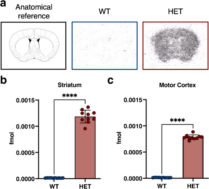
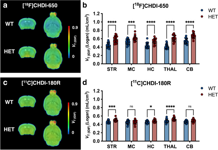
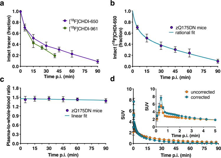
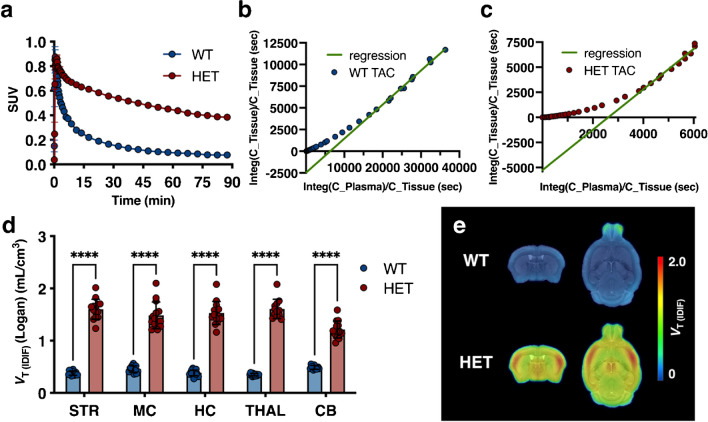
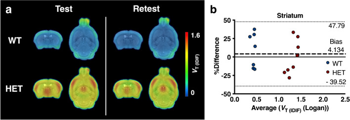
Plasma and brain radiometabolite profiles indicated a suitable metabolic profile for in vivo imaging of [F]CHDI-650. Both in vitro autoradiography and in vivo [F]CHDI-650 PET imaging at 9 months of age demonstrated a significant genotype effect (p < 0.0001) despite the poor test-retest reliability. [F]CHDI-650 PET imaging at 3 months of age displayed higher differentiation between genotypes when compared to [C]CHDI-180R.
Overall, [F]CHDI-650 allows for discrimination between HET and WT zQ175DN mice at 9 and 3 months of age. [F]CHDI-650 represents the first suitable F radioligand to image mHTT aggregates in mice and its clinical evaluation is underway.
正电子发射断层扫描(PET)对突变亨廷顿蛋白(mHTT)聚集体的成像,是监测亨廷顿病(HD)疾病进展以及候选治疗干预措施疗效的潜在工具。迄今为止,研究重点主要集中在 C 放射性配体的研究上;然而,有利的 F 放射性示踪剂将促进未来的临床转化。本工作旨在通过在 HD 的杂合子(HET)zQ175DN 小鼠模型中结合体内和体外方法,对新型 [F]CHDI-650 PET 放射性示踪剂进行特征描述。
在通过体外放射自显影对 [F]CHDI-650 进行特征描述后,我们在 9 月龄的 HET zQ175DN 小鼠模型和野生型(WT)同窝仔鼠中通过动态 PET 成像评估了体内血浆和脑放射性示踪剂的稳定性和动力学。此外,我们在 3 月龄时与之前发表的 [C]CHDI-180R 放射性配体进行了一项头对头比较研究。
血浆和脑放射性代谢物谱表明 [F]CHDI-650 具有适合体内成像的代谢特征。尽管测试-重测的可靠性较差,但体外放射自显影和 9 月龄时的体内 [F]CHDI-650 PET 成像均显示出显著的基因型效应(p < 0.0001)。与 [C]CHDI-180R 相比,3 月龄时的 [F]CHDI-650 PET 成像显示出更高的基因型差异。
总体而言,[F]CHDI-650 允许在 9 和 3 月龄时区分 HET 和 WT zQ175DN 小鼠。[F]CHDI-650 是第一个适合在小鼠中成像 mHTT 聚集体的 F 放射性配体,其临床评估正在进行中。