Molecular Imaging Center Antwerp (MICA), University of Antwerp, Universiteitsplein 1, Wilrijk, Antwerp, Belgium.
μNEURO Research Centre of Excellence, University of Antwerp, Antwerp, Belgium.
Eur J Nucl Med Mol Imaging. 2022 Mar;49(4):1166-1175. doi: 10.1007/s00259-021-05578-8. Epub 2021 Oct 15.
As several therapies aimed at lowering mutant huntingtin (mHTT) brain levels in Huntington's disease (HD) are currently being investigated, noninvasive positron emission tomography (PET) imaging of mHTT could be utilized to directly evaluate therapeutic efficacy and monitor disease progression. Here we characterized and longitudinally assessed the novel radioligand [C]CHDI-626 for mHTT PET imaging in the zQ175DN mouse model of HD.
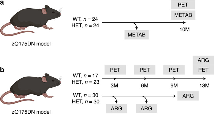
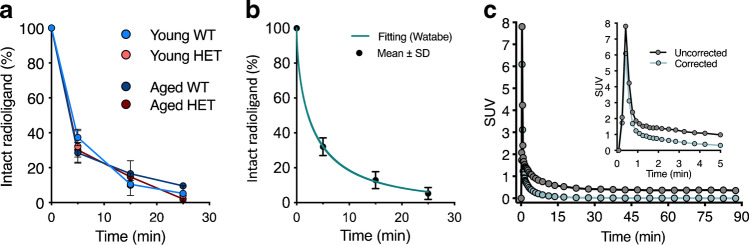
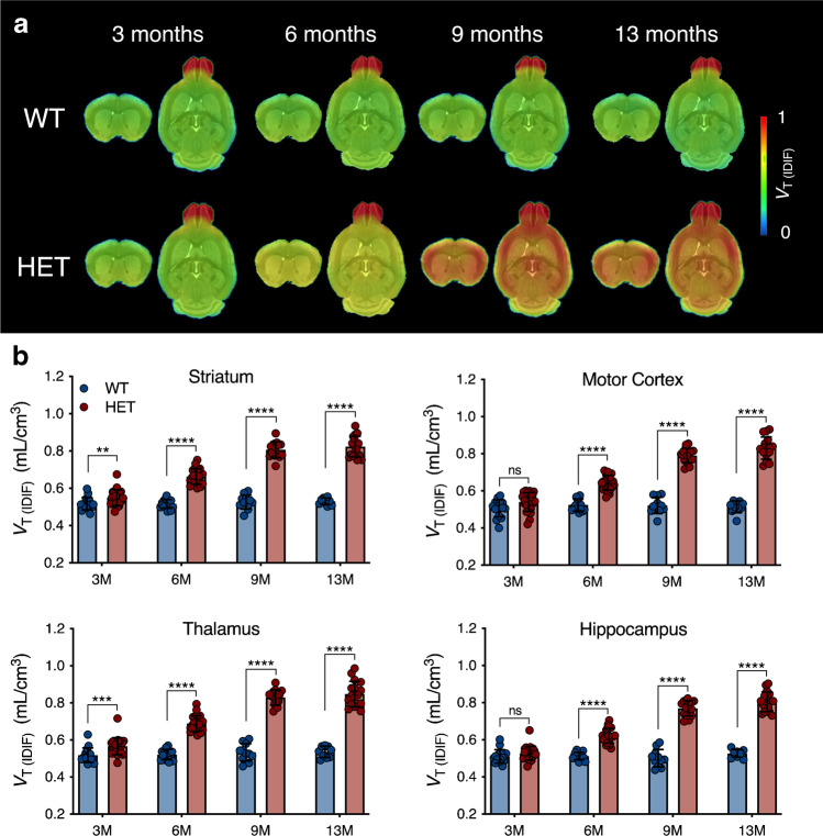
After evaluating radiometabolites and radioligand kinetics, we conducted longitudinal dynamic PET imaging at 3, 6, 9, and 13 months of age (M) in wild-type (WT, n = 17) and heterozygous (HET, n = 23) zQ175DN mice. Statistical analysis was performed to evaluate temporal and genotypic differences. Cross-sectional cohorts at each longitudinal time point were included for post-mortem [H]CHDI-626 autoradiography.
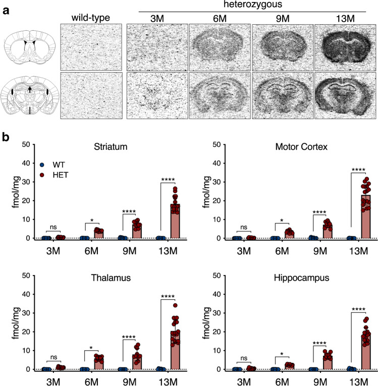
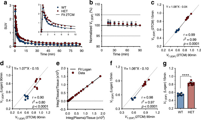
Despite fast metabolism and kinetics, the radioligand was suitable for PET imaging of mHTT. Longitudinal quantification could discriminate between genotypes already at premanifest stage (3 M), showing an age-associated increase in signal in HET mice in parallel with mHTT aggregate load progression, as supported by the post-mortem [H]CHDI-626 autoradiography.
With clinical evaluation underway, [C]CHDI-626 PET imaging appears to be a suitable preclinical candidate marker to monitor natural HD progression and for the evaluation of mHTT-lowering therapies.
由于目前正在研究几种旨在降低亨廷顿病(HD)中突变型亨廷顿蛋白(mHTT)脑水平的疗法,因此可以使用非侵入性正电子发射断层扫描(PET)成像来直接评估治疗效果并监测疾病进展。在这里,我们对新型放射性配体 [C]CHDI-626 进行了表征,并对 zQ175DN 小鼠 HD 模型进行了纵向评估。
在评估放射性代谢产物和放射性配体动力学后,我们在 3、6、9 和 13 个月龄(M)对野生型(WT,n=17)和杂合型(HET,n=23)zQ175DN 小鼠进行了纵向动态 PET 成像。进行了统计学分析以评估时间和基因型差异。每个纵向时间点的横截面队列都包括用于死后 [H]CHDI-626 放射自显影的分析。
尽管代谢和动力学较快,但该放射性配体仍适用于 mHTT 的 PET 成像。纵向定量分析已经可以在早期(3 个月)区分基因型,显示 HET 小鼠的信号随年龄呈上升趋势,与 mHTT 聚集体负荷进展平行,这得到了死后 [H]CHDI-626 放射自显影的支持。
随着临床评估的进行,[C]CHDI-626 PET 成像似乎是一种合适的临床前候选标志物,可用于监测自然发生的 HD 进展和评估 mHTT 降低疗法。