• 文献检索
  • 文档翻译
  • 深度研究
  • 学术资讯
  • Suppr Zotero 插件Zotero 插件
  • 邀请有礼
  • 套餐&价格
  • 历史记录
应用&插件
Suppr Zotero 插件Zotero 插件浏览器插件Mac 客户端Windows 客户端微信小程序
定价
高级版会员购买积分包购买API积分包
服务
文献检索文档翻译深度研究API 文档MCP 服务
关于我们
关于 Suppr公司介绍联系我们用户协议隐私条款
关注我们

Suppr 超能文献

核心技术专利:CN118964589B侵权必究
粤ICP备2023148730 号-1Suppr @ 2026

文献检索

告别复杂PubMed语法,用中文像聊天一样搜索,搜遍4000万医学文献。AI智能推荐,让科研检索更轻松。

立即免费搜索

文件翻译

保留排版,准确专业,支持PDF/Word/PPT等文件格式,支持 12+语言互译。

免费翻译文档

深度研究

AI帮你快速写综述,25分钟生成高质量综述,智能提取关键信息,辅助科研写作。

立即免费体验

常规和斑点追踪超声心动图变量在未排斥心脏移植患者与健康个体之间的比较分析。

Comparative Analysis of Conventional and Speckle Tracking Echocardiographic Variables between Patients with Unrejected Heart Transplants and Healthy Individuals.

机构信息

Universidade de Brasilia, Brasília, DF - Brasil.

Instituto de Cardiologia e Transplantes do Distrito Federal (ICTDF) , Brasília, DF - Brasil.

出版信息

Arq Bras Cardiol. 2024 Jul;121(8):e20230681. doi: 10.36660/abc.20230681.

DOI:10.36660/abc.20230681
PMID:39194042
原文链接:https://pmc.ncbi.nlm.nih.gov/articles/PMC11463340/
Abstract

BACKGROUND

Echocardiography is essential for the assessment of patients with heart transplants. However, normal values in such individuals are not clearly defined.

OBJECTIVES

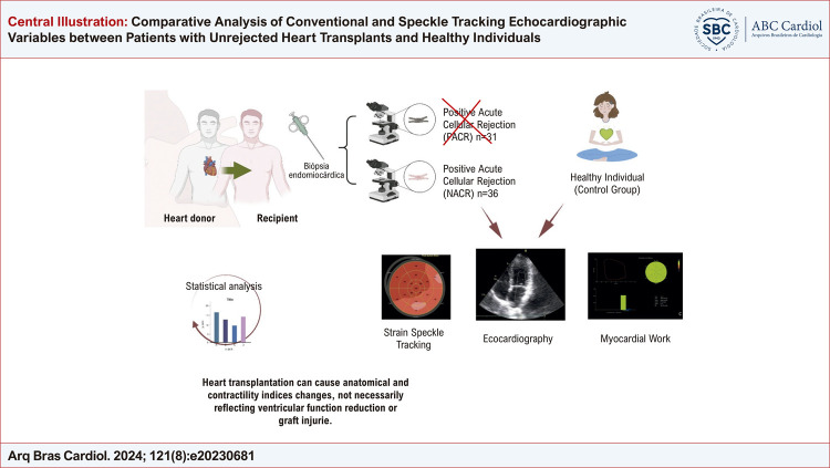
To compare conventional echocardiographic and speckle tracking variables between patients with unrejected heart transplants and healthy individuals.

METHODS

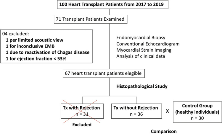
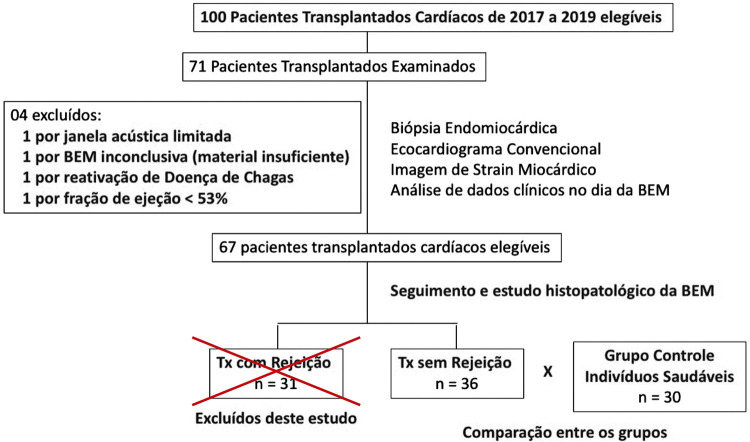
:A prospective study was conducted with adult patients having undergone heart transplantation at least one year earlier and submitted to endomyocardial biopsy followed by transthoracic echocardiogram (TTE). Conventional TTE measures and mechanical heart strain assessments using speckle tracking were performed and the results were compared to those of a group of healthy volunteers. Statistical significance was set at 5% (p < 0.05).

RESULTS

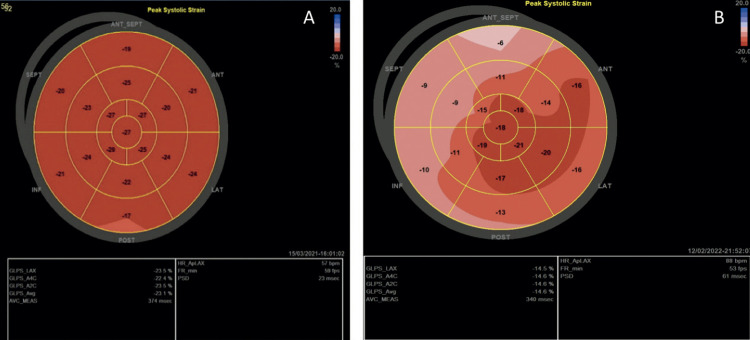
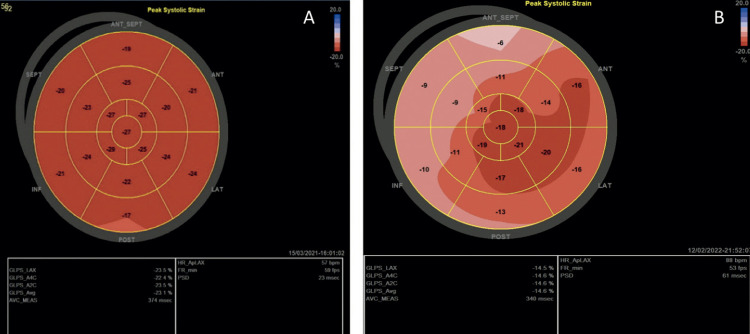
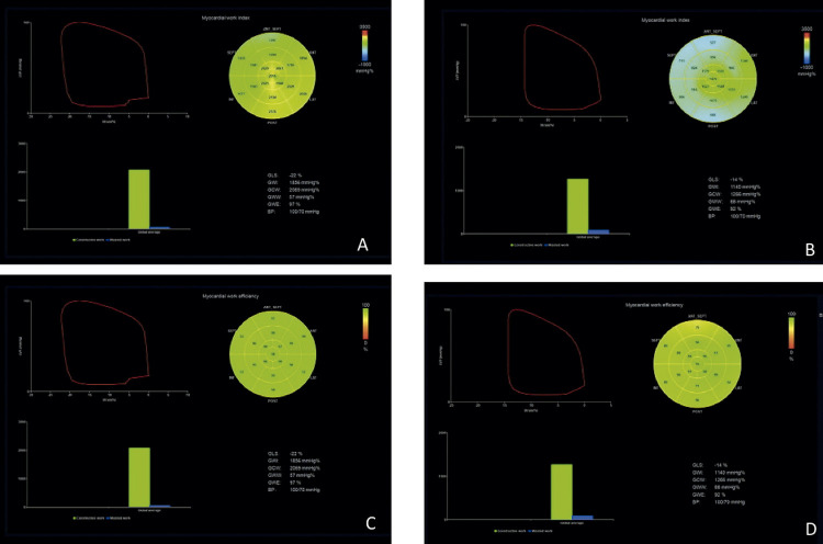
Thirty-six transplant patients without rejection were analyzed and compared to 30 healthy individuals. Chagas disease was the main reason for transplantation. Lower left ventricular global longitudinal strain expressed in absolute values was found (11.99% in transplant patients vs. 20.60% in controls; p <0.0001), right ventricular free wall longitudinal strain (16.67% in transplant patients vs. 25.50% in controls; p <0.0001) and myocardial work indices (p < 0.0001) as well as a larger size of the left atrium (38.17 ml/m2 in transplant patients vs. 18.98 ml/m2 in controls; p <0.0001) and greater mass and relative wall thickness (p <0.0001).

CONCLUSION

Stable patients having undergone heart transplants without rejection have differences concerning echocardiographic variables compared to healthy individuals. These findings indicate that conventional echocardiographic measures and heart mechanics are altered in transplant patients even in the absence of rejection. Such findings are relevant to the clinical context and follow-up of the patient.

摘要

背景

超声心动图对于评估心脏移植患者至关重要。然而,此类人群的正常值尚未明确界定。

目的

比较未排斥心脏移植患者与健康个体的常规超声心动图和斑点追踪变量。

方法

进行了一项前瞻性研究,纳入至少一年前接受过心脏移植且随后接受心肌活检和经胸超声心动图(TTE)的成年患者。进行了常规 TTE 测量和使用斑点追踪的机械心脏应变评估,并将结果与一组健康志愿者进行比较。统计显著性设为 5%(p<0.05)。

结果

分析了 36 例无排斥反应的移植患者,并与 30 名健康个体进行比较。Chagas 病是移植的主要原因。发现绝对值较低的左心室整体纵向应变(移植患者 11.99%,对照组 20.60%;p<0.0001)、右心室游离壁纵向应变(移植患者 16.67%,对照组 25.50%;p<0.0001)和心肌做功指数(p<0.0001)以及左心房较大的容积(移植患者 38.17ml/m2,对照组 18.98ml/m2;p<0.0001)和更大的质量和相对壁厚度(p<0.0001)。

结论

无排斥反应的心脏移植稳定患者的超声心动图变量与健康个体存在差异。这些发现表明,即使没有排斥反应,移植患者的常规超声心动图测量和心脏力学也会发生改变。这些发现与患者的临床情况和随访相关。

https://cdn.ncbi.nlm.nih.gov/pmc/blobs/8e7d/11463340/443585c360d2/0066-782X-abc-121-8-e20230681-gf04-en.jpg
https://cdn.ncbi.nlm.nih.gov/pmc/blobs/8e7d/11463340/ffe66f9ec59c/0066-782X-abc-121-8-e20230681-gf01.jpg
https://cdn.ncbi.nlm.nih.gov/pmc/blobs/8e7d/11463340/045816da1e10/0066-782X-abc-121-8-e20230681-gf02.jpg
https://cdn.ncbi.nlm.nih.gov/pmc/blobs/8e7d/11463340/821d5ec1a8a8/0066-782X-abc-121-8-e20230681-gf03.jpg
https://cdn.ncbi.nlm.nih.gov/pmc/blobs/8e7d/11463340/52323ef44c19/0066-782X-abc-121-8-e20230681-gf04.jpg
https://cdn.ncbi.nlm.nih.gov/pmc/blobs/8e7d/11463340/5bb21fa018e3/0066-782X-abc-121-8-e20230681-gf01-en.jpg
https://cdn.ncbi.nlm.nih.gov/pmc/blobs/8e7d/11463340/2945cd8e7fb2/0066-782X-abc-121-8-e20230681-gf02-en.jpg
https://cdn.ncbi.nlm.nih.gov/pmc/blobs/8e7d/11463340/127d8cdfd2d6/0066-782X-abc-121-8-e20230681-gf03-en.jpg
https://cdn.ncbi.nlm.nih.gov/pmc/blobs/8e7d/11463340/443585c360d2/0066-782X-abc-121-8-e20230681-gf04-en.jpg

相似文献

1
Comparative Analysis of Conventional and Speckle Tracking Echocardiographic Variables between Patients with Unrejected Heart Transplants and Healthy Individuals.常规和斑点追踪超声心动图变量在未排斥心脏移植患者与健康个体之间的比较分析。
Arq Bras Cardiol. 2024 Jul;121(8):e20230681. doi: 10.36660/abc.20230681.
2
Usefulness of speckle tracking echocardiography and biomarkers for detecting acute cellular rejection after heart transplantation.斑点追踪超声心动图和生物标志物在心脏移植后急性细胞排斥反应检测中的应用。
Cardiovasc Ultrasound. 2021 Jan 9;19(1):6. doi: 10.1186/s12947-020-00235-w.
3
Two-dimensional speckle-tracking strain echocardiography in long-term heart transplant patients: a study comparing deformation parameters and ejection fraction derived from echocardiography and multislice computed tomography.长期心脏移植患者的二维斑点追踪应变超声心动图:一项比较超声心动图和多层计算机断层扫描得出的变形参数及射血分数的研究
Eur J Echocardiogr. 2011 Jul;12(7):490-6. doi: 10.1093/ejechocard/jer064. Epub 2011 Jun 2.
4
Normal Reference Ranges for Transthoracic Echocardiography Following Heart Transplantation.心脏移植术后经胸超声心动图的正常参考范围。
J Am Soc Echocardiogr. 2018 Mar;31(3):349-360. doi: 10.1016/j.echo.2017.11.003. Epub 2017 Dec 21.
5
Usefulness of Two-Dimensional Strain Parameters to Diagnose Acute Rejection after Heart Transplantation.二维应变参数在诊断心脏移植术后急性排斥反应中的作用。
J Am Soc Echocardiogr. 2015 Oct;28(10):1149-56. doi: 10.1016/j.echo.2015.06.005. Epub 2015 Jul 10.
6
Evaluation of myocardial mechanics with three-dimensional speckle tracking echocardiography in heart transplant recipients: comparison with two-dimensional speckle tracking and relationship with clinical variables.三维斑点追踪超声心动图评价心脏移植受者心肌力学:与二维斑点追踪的比较及与临床变量的关系。
Eur Heart J Cardiovasc Imaging. 2013 Dec;14(12):1167-73. doi: 10.1093/ehjci/jet065. Epub 2013 Apr 23.
7
Myocardial strain and strain rate from speckle-tracking echocardiography are unable to differentiate asymptomatic biopsy-proven cellular rejection in the first year after cardiac transplantation.心脏移植术后第一年,斑点追踪超声心动图测量的心肌应变及应变率无法鉴别经活检证实的无症状细胞性排斥反应。
J Am Soc Echocardiogr. 2015 Apr;28(4):478-85. doi: 10.1016/j.echo.2014.12.013. Epub 2015 Jan 23.
8
Three-Dimensional Speckle-Tracking Echocardiographic Monitoring of Acute Rejection in Heart Transplant Recipients.心脏移植受者急性排斥反应的三维斑点追踪超声心动图监测
J Ultrasound Med. 2016 Jun;35(6):1167-76. doi: 10.7863/ultra.15.07013. Epub 2016 Apr 22.
9
Heart transplantation ten-year follow-ups: Deformation differentiation comparison of myocardial performance in left ventricle and right ventricle.心脏移植十年随访:左心室和右心室心肌性能的变形分化比较。
Clin Physiol Funct Imaging. 2020 Nov;40(6):415-422. doi: 10.1111/cpf.12660. Epub 2020 Sep 18.
10
Use of speckle-tracking echocardiography-derived strain and systolic strain rate measurements to predict rejection in transplant hearts with preserved ejection fraction.应用斑点追踪超声心动图应变和收缩期应变率测量来预测射血分数保留的移植心脏排斥反应。
BMC Cardiovasc Disord. 2018 Dec 22;18(1):241. doi: 10.1186/s12872-018-0980-4.

引用本文的文献

1
Do We Need New Echocardiographic Parameters for Transplant Recipients without Rejection?对于没有排斥反应的移植受者,我们是否需要新的超声心动图参数?
Arq Bras Cardiol. 2024 Sep 23;121(8):e20240452. doi: 10.36660/abc.20240452. eCollection 2024.

本文引用的文献

1
Position Statement on the Use of Myocardial Strain in Cardiology Routines by the Brazilian Society of Cardiology's Department Of Cardiovascular Imaging - 2023.《关于心肌应变在心血管影像学分会常规心脏病学中的应用的立场声明——2023》。
Arq Bras Cardiol. 2023 Dec;120(12):e20230646. doi: 10.36660/abc.20230646.
2
Acute Cellular Rejection in Heart Transplant Patients: Insights of Global Longitudinal Strain, Myocardial Work, and an Exclusive Group of Chagas Disease.心脏移植患者的急性细胞排斥反应:整体纵向应变、心肌做功及恰加斯病特定群体的相关见解
Front Cardiovasc Med. 2022 Apr 27;9:841698. doi: 10.3389/fcvm.2022.841698. eCollection 2022.
3
Longitudinal strain is an independent predictor of survival and response to therapy in patients with systemic AL amyloidosis.
纵向应变是系统性 AL 淀粉样变性患者生存和治疗反应的独立预测因子。
Eur Heart J. 2022 Jan 31;43(4):333-341. doi: 10.1093/eurheartj/ehab507.
4
The Utility of Myocardial Work in Clinical Practice.心肌做功在临床实践中的应用。
J Am Soc Echocardiogr. 2021 Aug;34(8):807-818. doi: 10.1016/j.echo.2021.04.013. Epub 2021 Apr 22.
5
Usefulness of speckle tracking echocardiography and biomarkers for detecting acute cellular rejection after heart transplantation.斑点追踪超声心动图和生物标志物在心脏移植后急性细胞排斥反应检测中的应用。
Cardiovasc Ultrasound. 2021 Jan 9;19(1):6. doi: 10.1186/s12947-020-00235-w.
6
Echocardiographic reference ranges for normal non-invasive myocardial work indices: results from the EACVI NORRE study.超声心动图正常无创心肌做功指数参考范围:EACVI NORRE 研究结果。
Eur Heart J Cardiovasc Imaging. 2019 May 1;20(5):582-590. doi: 10.1093/ehjci/jey188.
7
[Not Available].[无可用内容]
Arq Bras Cardiol. 2018 Aug;111(2):230-289. doi: 10.5935/abc.20180153.
8
Normal Reference Ranges for Transthoracic Echocardiography Following Heart Transplantation.心脏移植术后经胸超声心动图的正常参考范围。
J Am Soc Echocardiogr. 2018 Mar;31(3):349-360. doi: 10.1016/j.echo.2017.11.003. Epub 2017 Dec 21.
9
Recommendations for the Evaluation of Left Ventricular Diastolic Function by Echocardiography: An Update from the American Society of Echocardiography and the European Association of Cardiovascular Imaging.超声心动图评估左心室舒张功能的建议:美国超声心动图学会和欧洲心血管影像学会的更新
J Am Soc Echocardiogr. 2016 Apr;29(4):277-314. doi: 10.1016/j.echo.2016.01.011.
10
European Association of Cardiovascular Imaging/Cardiovascular Imaging Department of the Brazilian Society of Cardiology recommendations for the use of cardiac imaging to assess and follow patients after heart transplantation.欧洲心血管影像协会/巴西心脏病学会心血管影像学部关于心脏移植后评估和随访患者使用心脏影像的建议。
Eur Heart J Cardiovasc Imaging. 2015 Sep;16(9):919-48. doi: 10.1093/ehjci/jev139. Epub 2015 Jul 2.