Giusti Veronica, Miserocchi Giacomo, Sbanchi Giulia, Pannella Micaela, Hattinger Claudia Maria, Cesari Marilena, Fantoni Leonardo, Guerrieri Ania Naila, Bellotti Chiara, De Vita Alessandro, Spadazzi Chiara, Donati Davide Maria, Torsello Monica, Lucarelli Enrico, Ibrahim Toni, Mercatali Laura
Osteoncology, Bone and Soft Tissue Sarcomas and Innovative Therapies Unit, IRCCS Istituto Ortopedico Rizzoli, 40136 Bologna, Italy.
Preclinic and Osteoncology Unit, Biosciences Laboratory, IRCCS Istituto Romagnolo per lo Studio dei Tumori (IRST) "Dino Amadori", 47014 Meldola, Italy.
Biomedicines. 2024 Aug 21;12(8):1921. doi: 10.3390/biomedicines12081921.
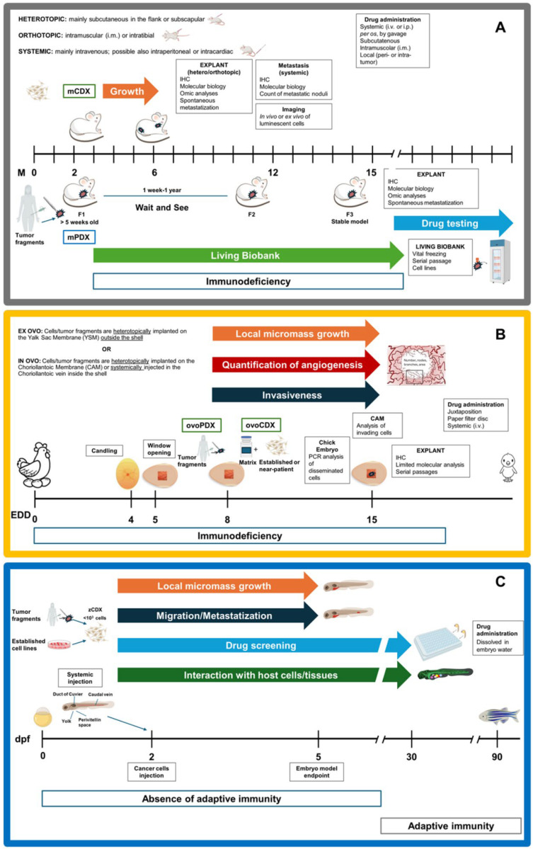
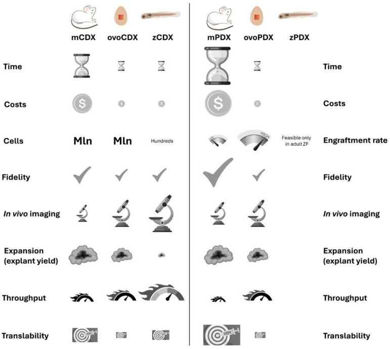
Musculoskeletal sarcomas pose major challenges to researchers and clinicians due to their rarity and heterogeneity. Xenografting human cells or tumor fragments in rodents is a mainstay for the generation of cancer models and for the preclinical trial of novel drugs. Lately, though, technical, intrinsic and ethical concerns together with stricter regulations have significantly curbed the employment of murine patient-derived xenografts (mPDX). In alternatives to murine PDXs, researchers have focused on embryonal systems such as chorioallantoic membrane (CAM) and zebrafish embryos. These systems are time- and cost-effective hosts for tumor fragments and near-patient cells. The CAM of the chick embryo represents a unique vascularized environment to host xenografts with high engraftment rates, allowing for ease of visualization and molecular detection of metastatic cells. Thanks to the transparency of the larvae, zebrafish allow for the tracking of tumor development and metastatization, enabling high-throughput drug screening. This review will focus on xenograft models of musculoskeletal sarcomas to highlight the intrinsic and technically distinctive features of the different hosts, and how they can be exploited to elucidate biological mechanisms beneath the different phases of the tumor's natural history and in drug development. Ultimately, the review suggests the combination of different models as an advantageous approach to boost basic and translational research.
肌肉骨骼肉瘤因其罕见性和异质性,给研究人员和临床医生带来了重大挑战。将人类细胞或肿瘤片段移植到啮齿动物体内,是建立癌症模型和开展新型药物临床前试验的主要方法。然而,近年来,技术、内在因素和伦理问题,以及更严格的法规,显著限制了小鼠患者来源异种移植模型(mPDX)的应用。在小鼠PDX模型的替代方案中,研究人员将重点放在了胚胎系统上,如鸡胚绒毛尿囊膜(CAM)和斑马鱼胚胎。这些系统对于肿瘤片段和类患者细胞而言,是具有时间和成本效益的宿主。鸡胚的CAM代表了一种独特的血管化环境,能够以高移植率宿主异种移植物,便于观察转移细胞并进行分子检测。由于斑马鱼幼虫的透明性,其可用于追踪肿瘤的发展和转移,从而实现高通量药物筛选。本综述将聚焦于肌肉骨骼肉瘤的异种移植模型,以突出不同宿主的内在和技术上的独特特征,以及如何利用它们来阐明肿瘤自然史不同阶段和药物开发背后的生物学机制。最终,本综述建议将不同模型结合起来,作为推动基础研究和转化研究的一种有利方法。