• 文献检索
  • 文档翻译
  • 深度研究
  • 学术资讯
  • Suppr Zotero 插件Zotero 插件
  • 邀请有礼
  • 套餐&价格
  • 历史记录
应用&插件
Suppr Zotero 插件Zotero 插件浏览器插件Mac 客户端Windows 客户端微信小程序
定价
高级版会员购买积分包购买API积分包
服务
文献检索文档翻译深度研究API 文档MCP 服务
关于我们
关于 Suppr公司介绍联系我们用户协议隐私条款
关注我们

Suppr 超能文献

核心技术专利:CN118964589B侵权必究
粤ICP备2023148730 号-1Suppr @ 2026

文献检索

告别复杂PubMed语法,用中文像聊天一样搜索,搜遍4000万医学文献。AI智能推荐,让科研检索更轻松。

立即免费搜索

文件翻译

保留排版,准确专业,支持PDF/Word/PPT等文件格式,支持 12+语言互译。

免费翻译文档

深度研究

AI帮你快速写综述,25分钟生成高质量综述,智能提取关键信息,辅助科研写作。

立即免费体验

使用高频超声引导美容填充剂注射的最佳实践——第1部分:面部上三分之一区域

Best Practices for the Use of High-Frequency Ultrasound to Guide Aesthetic Filler Injections-Part 1: Upper Third of the Face.

作者信息

Sigrist Rosa, Desyatnikova Stella, Chammas Maria Cristina, Vasconcelos-Berg Roberta

机构信息

Department of Radiology, School of Medicine, University of São Paulo, São Paulo 05403-010, Brazil.

The Stella Center for Facial Plastic Surgery, Seattle, WA 98101, USA.

出版信息

Diagnostics (Basel). 2024 Aug 8;14(16):1718. doi: 10.3390/diagnostics14161718.

DOI:10.3390/diagnostics14161718
PMID:39202206
原文链接:https://pmc.ncbi.nlm.nih.gov/articles/PMC11353849/
Abstract

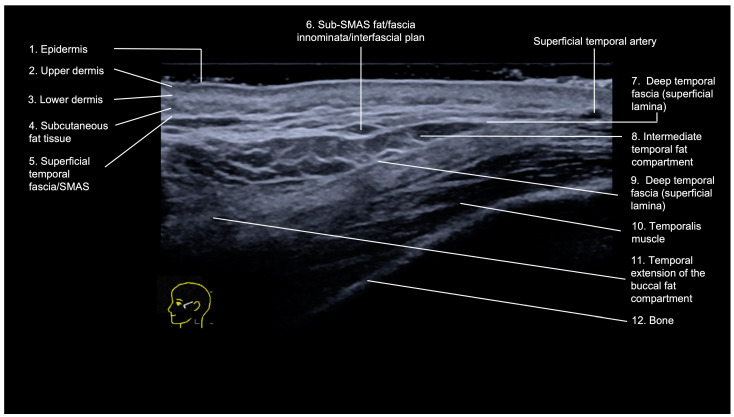
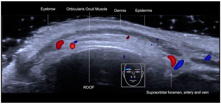
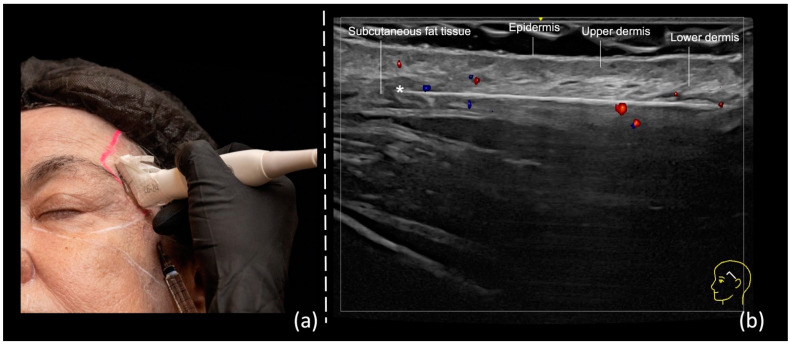
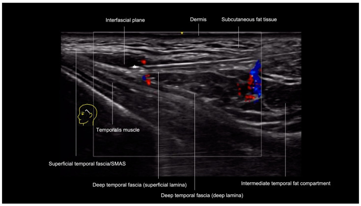
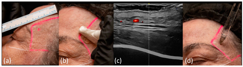
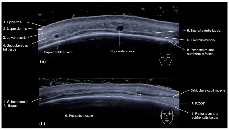
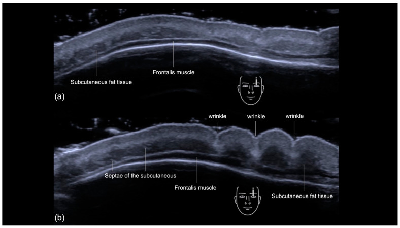
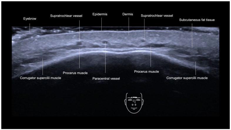
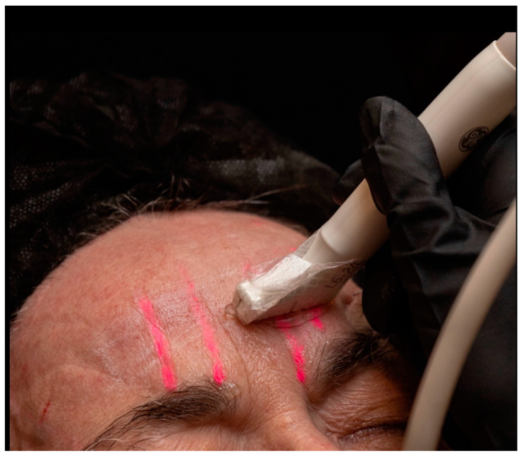
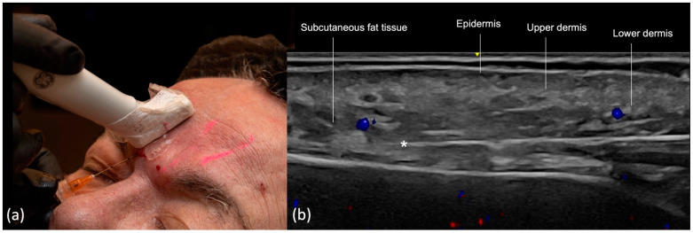
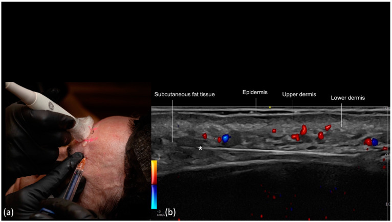
Filler injections in the upper face pose significant challenges due to its complex anatomy and proximity to vascular structures. High-frequency Doppler ultrasound offers real-time visualization of facial anatomy, improving both safety and aesthetic outcomes. This paper presents a detailed overview of the ultrasonographic anatomy of the temples, forehead, and glabella, along with reproducible, ultrasound-guided filler injection techniques for these areas. We use two scanning techniques previously described: "scan before injecting" and "scan while injecting", applicable to subdermal, interfascial, and supraperiosteal planes in the temporal region, as well as the glabella, forehead, and supraorbital region. Ultrasound guidance for filler injections in the upper face can enhance procedural efficacy and safety. By integrating real-time imaging, practitioners can navigate the intricate vascular anatomy more effectively, thereby minimizing the risk of complications. This study highlights the need for ongoing research and continuous education to further refine these techniques and improve patient outcomes.

摘要

由于上半面部解剖结构复杂且靠近血管结构,在上半面部进行填充剂注射面临重大挑战。高频多普勒超声可实时显示面部解剖结构,提高安全性和美学效果。本文详细概述了颞部、额部和眉间的超声解剖结构,以及这些区域可重复的超声引导下填充剂注射技术。我们采用了之前描述的两种扫描技术:“注射前扫描”和“注射时扫描”,适用于颞部区域以及眉间、额部和眶上区域的皮下、筋膜间和骨膜上平面。上半面部填充剂注射的超声引导可提高操作的有效性和安全性。通过整合实时成像,从业者可以更有效地避开复杂的血管解剖结构,从而将并发症风险降至最低。本研究强调了持续研究和持续教育的必要性,以进一步完善这些技术并改善患者预后。

https://cdn.ncbi.nlm.nih.gov/pmc/blobs/435c/11353849/18d3ecf5aec9/diagnostics-14-01718-g011.jpg
https://cdn.ncbi.nlm.nih.gov/pmc/blobs/435c/11353849/854d97c8c8f8/diagnostics-14-01718-g001.jpg
https://cdn.ncbi.nlm.nih.gov/pmc/blobs/435c/11353849/3ca357c33bf8/diagnostics-14-01718-g002.jpg
https://cdn.ncbi.nlm.nih.gov/pmc/blobs/435c/11353849/4a1f22afb2ce/diagnostics-14-01718-g003.jpg
https://cdn.ncbi.nlm.nih.gov/pmc/blobs/435c/11353849/de76c2f05cbf/diagnostics-14-01718-g004.jpg
https://cdn.ncbi.nlm.nih.gov/pmc/blobs/435c/11353849/7aacee0bb8b5/diagnostics-14-01718-g005.jpg
https://cdn.ncbi.nlm.nih.gov/pmc/blobs/435c/11353849/3b84bb818293/diagnostics-14-01718-g006.jpg
https://cdn.ncbi.nlm.nih.gov/pmc/blobs/435c/11353849/a72fa13cb57f/diagnostics-14-01718-g007.jpg
https://cdn.ncbi.nlm.nih.gov/pmc/blobs/435c/11353849/8977dd92cc09/diagnostics-14-01718-g008.jpg
https://cdn.ncbi.nlm.nih.gov/pmc/blobs/435c/11353849/5acdff20faf2/diagnostics-14-01718-g009.jpg
https://cdn.ncbi.nlm.nih.gov/pmc/blobs/435c/11353849/c52bf9615c09/diagnostics-14-01718-g010.jpg
https://cdn.ncbi.nlm.nih.gov/pmc/blobs/435c/11353849/18d3ecf5aec9/diagnostics-14-01718-g011.jpg

相似文献

1
Best Practices for the Use of High-Frequency Ultrasound to Guide Aesthetic Filler Injections-Part 1: Upper Third of the Face.使用高频超声引导美容填充剂注射的最佳实践——第1部分:面部上三分之一区域
Diagnostics (Basel). 2024 Aug 8;14(16):1718. doi: 10.3390/diagnostics14161718.
2
Hyaluronic Acid Filler Injection Guided by Doppler Ultrasound.多普勒超声引导下透明质酸填充剂注射
Arch Plast Surg. 2023 Aug 2;50(4):348-353. doi: 10.1055/s-0043-1770078. eCollection 2023 Jul.
3
Ultrasound-Guided Temple Filler Injection.超声引导太阳穴填充注射。
Facial Plast Surg Aesthet Med. 2022 Nov-Dec;24(6):501-503. doi: 10.1089/fpsam.2022.0176.
4
Doppler Ultrasound-Guided Filler Injections: Useful Tips to Integrate Ultrasound in Daily Practice.超声引导下填充剂注射:将超声整合到日常实践中的有用技巧。
Aesthet Surg J. 2023 Jun 14;43(7):773-783. doi: 10.1093/asj/sjac353.
5
A roadmap for safety during facial filler injections: A fresh frozen cadaver study.面部填充物注射安全路线图:新鲜冷冻尸体研究。
J Plast Reconstr Aesthet Surg. 2023 Nov;86:155-164. doi: 10.1016/j.bjps.2023.06.029. Epub 2023 Jun 13.
6
Description of a safe doppler ultrasound-guided technique for hyaluronic acid filler in the face-A method to avoid adverse vascular events.一种用于面部透明质酸填充剂的安全多普勒超声引导技术的描述——一种避免不良血管事件的方法。
J Cosmet Dermatol. 2022 Jul;21(7):2783-2787. doi: 10.1111/jocd.14492. Epub 2021 Sep 29.
7
The six different injection techniques for the temple relevant for soft tissue filler augmentation procedures - Clinical anatomy and danger zones.与软组织填充剂隆颞手术相关的六种不同颞部注射技术——临床解剖与危险区域。
J Cosmet Dermatol. 2020 Jul;19(7):1570-1579. doi: 10.1111/jocd.13491. Epub 2020 Jun 1.
8
Pre-injection ultrasound scanning for treating temporal hollowing.超声预处理注射治疗颞部凹陷。
J Cosmet Dermatol. 2022 Jun;21(6):2420-2425. doi: 10.1111/jocd.14471. Epub 2021 Sep 24.
9
Pinch anatomy: An injection guide for temple filler injections.捏掐解剖:太阳穴填充物注射的注射指南。
Dermatol Ther. 2020 Nov;33(6):e13983. doi: 10.1111/dth.13983. Epub 2020 Jul 26.
10
Facial vascular danger zones for filler injections.面部血管危险区域填充注射。
Dermatol Ther. 2020 Nov;33(6):e14285. doi: 10.1111/dth.14285. Epub 2020 Sep 18.

引用本文的文献

1
Evaluation of Facial Artery Course Variations, Diameters, and Depth Using Doppler Ultrasonography: A Systematic Review and Meta-Analysis.使用多普勒超声评估面动脉走行变异、直径和深度:系统评价与荟萃分析
J Cosmet Dermatol. 2025 Sep;24(9):e70431. doi: 10.1111/jocd.70431.
2
The SIBUS-IN Fingerheld Ultrasound Machine.SIBUS-IN手持式超声诊断仪
J Cosmet Dermatol. 2025 Jul;24(7):e70313. doi: 10.1111/jocd.70313.
3
Best Practices for the Use of High-Frequency Ultrasound to Guide Esthetic Filler Injections-Part 3: Lower Third of the Face.

本文引用的文献

1
Increasing precision during neuromodulator injections for frontal rhytids-Using ultrasound imaging to identify the line of convergence.提高额部皱纹神经调节剂注射的精准度——应用超声成像技术确定汇聚线。
J Cosmet Dermatol. 2024 Jul;23(7):2373-2379. doi: 10.1111/jocd.16368. Epub 2024 May 16.
2
Response to: Demystifying the Forehead Using Ultrasonography.回应:《超声检查揭秘额头》
Aesthet Surg J. 2024 Jun 14;44(7):NP525-NP526. doi: 10.1093/asj/sjae087.
3
Demystifying the Forehead With Ultrasonography.用超声检查揭开额头之谜。
使用高频超声引导美容填充剂注射的最佳实践——第3部分:面部下三分之一区域
Diagnostics (Basel). 2025 Apr 2;15(7):921. doi: 10.3390/diagnostics15070921.
4
High-Frequency and Ultra-High-Frequency Ultrasound in Dermatologic Diseases and Aesthetic Medicine.高频和超高频超声在皮肤病及美容医学中的应用
Medicina (Kaunas). 2025 Jan 26;61(2):220. doi: 10.3390/medicina61020220.
Aesthet Surg J. 2024 Jun 14;44(7):NP523-NP524. doi: 10.1093/asj/sjae065.
4
Anatomy of the temporal region to guide filler injections.颞区解剖结构指导填充剂注射。
Surg Radiol Anat. 2024 May;46(5):615-624. doi: 10.1007/s00276-024-03340-x. Epub 2024 Mar 13.
5
An Exploration of the Anatomy of the Forehead of Asians and Its Relationship With Forehead Lines Based on Ultrasound Imaging.基于超声成像对亚洲人前额解剖结构及其与额纹关系的探索。
Aesthet Surg J. 2023 Oct 13;43(11):NP956-NP961. doi: 10.1093/asj/sjad133.
6
Doppler Ultrasound-Guided Filler Injections: Useful Tips to Integrate Ultrasound in Daily Practice.超声引导下填充剂注射:将超声整合到日常实践中的有用技巧。
Aesthet Surg J. 2023 Jun 14;43(7):773-783. doi: 10.1093/asj/sjac353.
7
Ultrasound as an Educational Tool in Facial Aesthetic Injections.超声作为面部美容注射的一种教育工具
Plast Reconstr Surg Glob Open. 2022 Dec 12;10(12):e4639. doi: 10.1097/GOX.0000000000004639. eCollection 2022 Dec.
8
Evaluation of Supratrochlear, Supraorbital and Angular Artery Course Variations and Depth by Doppler Ultrasound.通过多普勒超声评估滑车上动脉、眶上动脉和内眦动脉的走行变异及深度
Aesthetic Plast Surg. 2023 Apr;47(2):791-798. doi: 10.1007/s00266-022-03188-9. Epub 2022 Nov 28.
9
Ultrasound-Guided Temple Filler Injection.超声引导太阳穴填充注射。
Facial Plast Surg Aesthet Med. 2022 Nov-Dec;24(6):501-503. doi: 10.1089/fpsam.2022.0176.
10
Understanding the Anatomic Layers of the Forehead Seen on Ultrasound: A Guide for Injectable Fillers and Chemodenervation.了解超声下所见前额的解剖层次:注射填充剂和化学去神经支配指南
Facial Plast Surg Aesthet Med. 2023 Mar-Apr;25(2):179-181. doi: 10.1089/fpsam.2022.0130. Epub 2022 Sep 13.