• 文献检索
  • 文档翻译
  • 深度研究
  • 学术资讯
  • Suppr Zotero 插件Zotero 插件
  • 邀请有礼
  • 套餐&价格
  • 历史记录
应用&插件
Suppr Zotero 插件Zotero 插件浏览器插件Mac 客户端Windows 客户端微信小程序
定价
高级版会员购买积分包购买API积分包
服务
文献检索文档翻译深度研究API 文档MCP 服务
关于我们
关于 Suppr公司介绍联系我们用户协议隐私条款
关注我们

Suppr 超能文献

核心技术专利:CN118964589B侵权必究
粤ICP备2023148730 号-1Suppr @ 2026

文献检索

告别复杂PubMed语法,用中文像聊天一样搜索,搜遍4000万医学文献。AI智能推荐,让科研检索更轻松。

立即免费搜索

文件翻译

保留排版,准确专业,支持PDF/Word/PPT等文件格式,支持 12+语言互译。

免费翻译文档

深度研究

AI帮你快速写综述,25分钟生成高质量综述,智能提取关键信息,辅助科研写作。

立即免费体验

薄型非色素性和低色素性黑素瘤:临床病理和皮肤镜特征。

Thin Amelanotic and Hypomelanotic Melanoma: Clinicopathological and Dermoscopic Features.

机构信息

Unit of Dermatology and Cosmetology, IRCCS San Raffaele Scientific Institute, 20132 Milan, Italy.

La Sapienza University of Rome, 00185 Rome, Italy.

出版信息

Medicina (Kaunas). 2024 Jul 30;60(8):1239. doi: 10.3390/medicina60081239.

DOI:10.3390/medicina60081239
PMID:39202520
原文链接:https://pmc.ncbi.nlm.nih.gov/articles/PMC11356094/
Abstract

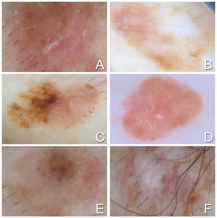
: Amelanotic/hypomelanotic melanomas (AHMs) account for 2-8% of all cutaneous melanomas. Due to their clinical appearance and the lack of specific dermoscopic indicators, AHMs are challenging to diagnose, particularly in thinner cutaneous lesions. The aim of our study was to evaluate the clinicopathological and dermoscopic features of thin AHMs. Identifying the baseline clinical-pathological features and dermoscopic aspects of thin AHMs is crucial to better understand this entity. : We divided the AHM cohort into two groups based on Breslow thickness: thin (≤1.00 mm) and thick (>1.00 mm). This stratification helped identify any significant clinicopathological differences between the groups. For dermoscopic analysis, we employed the "pattern analysis" approach, which involves a simultaneous and subjective assessment of different criteria. : Out of the 2.800 melanomas analyzed for Breslow thickness, 153 were identified as AHMs. Among these, 65 patients presented with thin AHMs and 88 with thick AHMs. Red hair color and phototype II were more prevalent in patients with thin AHMs. The trunk was the most common anatomic site for thin AHMs. Patients with thin AHMs showed a higher number of multiple melanomas. Dermoscopic analysis revealed no significant difference between thin AHMs and thick AHMs, except for a more frequent occurrence of residual reticulum in thin AHMs. : Thin AHMs typically affect individuals with lower phototypes and red hair color. These aspects can be related to the higher presence of pheomelanin, which provides limited protection against sun damage. This also correlates with the fact that the trunk, a site commonly exposed to intermittent sun exposure, is the primary anatomical location for thin AHMs. Multiple primary melanomas are more common in patients with thin AHMs, likely due to an intrinsic predisposition as well as greater periodic dermatologic follow-ups in this class of patients. Apart from the presence of residual reticulum, no other significant dermoscopic differences were observed, complicating the differential diagnosis between thin and thick AHMs based on dermoscopy alone.

摘要

无色素性/低色素性黑素瘤(AHM)占所有皮肤黑素瘤的 2-8%。由于其临床外观和缺乏特定的皮肤镜指标,AHM 的诊断具有挑战性,尤其是在较薄的皮肤病变中。我们研究的目的是评估薄的 AHM 的临床病理和皮肤镜特征。确定薄的 AHM 的基线临床病理特征和皮肤镜特征对于更好地理解这种实体至关重要。

我们根据 Breslow 厚度将 AHM 队列分为两组:薄(≤1.00mm)和厚(>1.00mm)。这种分层有助于确定两组之间是否存在任何显著的临床病理差异。对于皮肤镜分析,我们采用了“模式分析”方法,该方法涉及对不同标准进行同时和主观评估。

在分析 Breslow 厚度的 2800 个黑素瘤中,有 153 个被确定为 AHM。其中,65 例患者表现为薄的 AHM,88 例患者表现为厚的 AHM。薄的 AHM 患者中红发和 II 型光型更为常见。躯干是薄的 AHM 最常见的解剖部位。薄的 AHM 患者中多发性黑素瘤的数量较多。皮肤镜分析显示,除了薄的 AHM 中更频繁地出现残留网纹外,薄的 AHM 和厚的 AHM 之间没有明显差异。

薄的 AHM 通常影响光型和红发颜色较低的个体。这些方面可能与较高含量的 pheomelanin 有关,pheomelanin 对太阳损伤的保护作用有限。这也与躯干是薄的 AHM 的主要解剖部位的事实相关,躯干是一个经常间歇性暴露于阳光的部位。薄的 AHM 患者中多发性原发性黑素瘤更为常见,这可能是由于内在倾向以及此类患者更频繁的定期皮肤科随访所致。除了残留网纹的存在外,没有观察到其他明显的皮肤镜差异,这使得仅凭皮肤镜难以区分薄的和厚的 AHM。

https://cdn.ncbi.nlm.nih.gov/pmc/blobs/b43f/11356094/f8ba0835e2d5/medicina-60-01239-g001.jpg

相似文献

1
Thin Amelanotic and Hypomelanotic Melanoma: Clinicopathological and Dermoscopic Features.薄型非色素性和低色素性黑素瘤:临床病理和皮肤镜特征。
Medicina (Kaunas). 2024 Jul 30;60(8):1239. doi: 10.3390/medicina60081239.
2
Amelanotic/hypomelanotic melanoma: clinical and dermoscopic features.无色素性/色素减退性黑色素瘤:临床及皮肤镜特征
Br J Dermatol. 2004 Jun;150(6):1117-24. doi: 10.1111/j.1365-2133.2004.05928.x.
3
Clinicopathological, dermoscopic features and circumstances of diagnosis of amelanotic or hypomelanotic melanoma: A prospective multicentric study in the French private medical sector.无色素性或低色素性黑色素瘤的临床病理、皮肤镜特征及诊断情况:法国私立医疗领域的一项前瞻性多中心研究。
Ann Dermatol Venereol. 2024 Mar;151(1):103249. doi: 10.1016/j.annder.2024.103249. Epub 2024 Feb 29.
4
Clinicopathological and dermoscopic features of amelanotic and hypomelanotic melanoma: a retrospective multicentric study.无色素性和低色素性黑色素瘤的临床病理和皮肤镜特征:一项回顾性多中心研究。
Int J Dermatol. 2020 Nov;59(11):1371-1380. doi: 10.1111/ijd.15064. Epub 2020 Jul 29.
5
Clinically amelanotic or hypomelanotic melanoma: Anatomic distribution, risk factors, and survival.临床无色素性或低色素性黑色素瘤:解剖分布、危险因素和生存。
J Am Acad Dermatol. 2018 Oct;79(4):645-651.e4. doi: 10.1016/j.jaad.2018.04.045.
6
More than one shade of pink as a marker of early amelanotic/hypomelanotic melanoma.多种粉红色调提示早期无黑色素瘤/低黑色素瘤性黑素瘤。
J Dermatol. 2024 Jul;51(7):999-1003. doi: 10.1111/1346-8138.17200. Epub 2024 Mar 25.
7
Analysis of the dermoscopic features of excised melanomas and their relation with tumor thickness in a tertiary hospital in Brazil.巴西一家三级医院切除黑色素瘤的皮肤镜特征分析及其与肿瘤厚度的关系。
Int J Dermatol. 2024 Aug;63(8):1064-1070. doi: 10.1111/ijd.17047. Epub 2024 Feb 28.
8
Amelanotic/Hypomelanotic melanoma--is dermatoscopy useful for diagnosis?无色素性/低色素性黑素瘤——皮肤镜检查对诊断有用吗?
J Dtsch Dermatol Ges. 2003 May;1(5):369-73. doi: 10.1046/j.1610-0387.2003.02042.x.
9
Clinical and dermoscopic characteristics of amelanotic melanomas that are not of the nodular subtype.非结节型无黑色素瘤的临床和皮肤镜特征。
J Eur Acad Dermatol Venereol. 2012 May;26(5):591-6. doi: 10.1111/j.1468-3083.2011.04122.x. Epub 2011 May 18.
10
Dermoscopic evaluation of amelanotic and hypomelanotic melanoma.无色素性和色素减退性黑色素瘤的皮肤镜评估
Arch Dermatol. 2008 Sep;144(9):1120-7. doi: 10.1001/archderm.144.9.1120.

本文引用的文献

1
Malignant Melanoma: An Overview, New Perspectives, and Vitamin D Signaling.恶性黑色素瘤:概述、新观点及维生素D信号传导
Cancers (Basel). 2024 Jun 18;16(12):2262. doi: 10.3390/cancers16122262.
2
Dermoscopy and reflectance confocal microscopy of solitary flat pink lesions: A new combined score to diagnose amelanotic melanoma.孤立性扁平粉色皮损的皮肤镜检查和反射式共聚焦显微镜检查:一种诊断无色素性黑色素瘤的新综合评分法。
J Eur Acad Dermatol Venereol. 2025 Jan;39(1):109-116. doi: 10.1111/jdv.19991. Epub 2024 Apr 4.
3
A twenty year perspective on melanoma therapy.
对黑色素瘤治疗的二十年展望。
Pigment Cell Melanoma Res. 2023 Nov;36(6):563-575. doi: 10.1111/pcmr.13125. Epub 2023 Sep 28.
4
Cutaneous melanoma.皮肤黑素瘤。
Lancet. 2023 Aug 5;402(10400):485-502. doi: 10.1016/S0140-6736(23)00821-8. Epub 2023 Jul 24.
5
How cancer hijacks the body's homeostasis through the neuroendocrine system.癌症如何通过神经内分泌系统劫持身体的内稳态。
Trends Neurosci. 2023 Apr;46(4):263-275. doi: 10.1016/j.tins.2023.01.003. Epub 2023 Feb 17.
6
Melanoma, Melanin, and Melanogenesis: The Yin and Yang Relationship.黑色素瘤、黑色素与黑色素生成:阴阳关系
Front Oncol. 2022 Mar 14;12:842496. doi: 10.3389/fonc.2022.842496. eCollection 2022.
7
Recent Advances in the Treatment of Melanoma.黑色素瘤治疗的最新进展
N Engl J Med. 2021 Jun 10;384(23):2229-2240. doi: 10.1056/NEJMra2034861.
8
Melanization as unfavorable factor in amelanotic melanoma cell biology.黑色素化作为无黑色素性黑色素瘤细胞生物学中的不利因素。
Protoplasma. 2021 Sep;258(5):935-948. doi: 10.1007/s00709-021-01613-5. Epub 2021 Jan 27.
9
Clinicopathological and dermoscopic features of amelanotic and hypomelanotic melanoma: a retrospective multicentric study.无色素性和低色素性黑色素瘤的临床病理和皮肤镜特征:一项回顾性多中心研究。
Int J Dermatol. 2020 Nov;59(11):1371-1380. doi: 10.1111/ijd.15064. Epub 2020 Jul 29.
10
Protein-Bound Polysaccharides from Induce RIPK1/RIPK3/MLKL-Mediated Necroptosis in ER-Positive Breast Cancer and Amelanotic Melanoma Cells.从 中提取的蛋白结合多糖可诱导内质网阳性乳腺癌和无黑色素性黑色素瘤细胞中 RIPK1/RIPK3/MLKL 介导的坏死性凋亡。
Cell Physiol Biochem. 2020 Jun 13;54(4):591-604. doi: 10.33594/000000242.