• 文献检索
  • 文档翻译
  • 深度研究
  • 学术资讯
  • Suppr Zotero 插件Zotero 插件
  • 邀请有礼
  • 套餐&价格
  • 历史记录
应用&插件
Suppr Zotero 插件Zotero 插件浏览器插件Mac 客户端Windows 客户端微信小程序
定价
高级版会员购买积分包购买API积分包
服务
文献检索文档翻译深度研究API 文档MCP 服务
关于我们
关于 Suppr公司介绍联系我们用户协议隐私条款
关注我们

Suppr 超能文献

核心技术专利:CN118964589B侵权必究
粤ICP备2023148730 号-1Suppr @ 2026

文献检索

告别复杂PubMed语法,用中文像聊天一样搜索,搜遍4000万医学文献。AI智能推荐,让科研检索更轻松。

立即免费搜索

文件翻译

保留排版,准确专业,支持PDF/Word/PPT等文件格式,支持 12+语言互译。

免费翻译文档

深度研究

AI帮你快速写综述,25分钟生成高质量综述,智能提取关键信息,辅助科研写作。

立即免费体验

磁共振成像检测亨廷顿病的结构性脑变化:来自小鼠模型的数据综述。

Magnetic Resonance Imaging to Detect Structural Brain Changes in Huntington's Disease: A Review of Data from Mouse Models.

机构信息

Department of Chemistry, Memorial University of Newfoundland, St. John's, Newfoundland and Labrador, Canada.

Discipline of Radiology, Memorial University of Newfoundland, St. John's, Newfoundland and Labrador, Canada.

出版信息

J Huntingtons Dis. 2024;13(3):279-299. doi: 10.3233/JHD-240045.

DOI:10.3233/JHD-240045
PMID:39213087
原文链接:https://pmc.ncbi.nlm.nih.gov/articles/PMC11494634/
Abstract

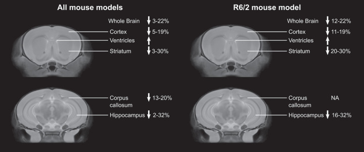
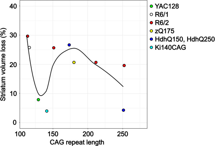
Structural magnetic resonance imaging (MRI) is a powerful tool to visualize 3D neuroanatomy and assess pathology and disease progression in neurodegenerative disorders such as Huntington's disease (HD). The development of mouse models of HD that reproduce many of the psychiatric, motor and cognitive impairments observed in human HD has improved our understanding of the disease and provided opportunities for testing novel therapies. Similar to the clinical scenario, MRI of mouse models of HD demonstrates onset and progression of brain pathology. Here, we provided an overview of the articles that used structural MRI in mouse models of HD to date, highlighting the differences between studies and models and describing gaps in the current state of knowledge and recommendations for future studies.

摘要

结构磁共振成像(MRI)是一种强大的工具,可用于可视化 3D 神经解剖结构,并评估神经退行性疾病(如亨廷顿病(HD))中的病理学和疾病进展。开发出能够重现人类 HD 中观察到的许多精神、运动和认知障碍的 HD 小鼠模型,这提高了我们对该疾病的认识,并为测试新疗法提供了机会。与临床情况类似,HD 小鼠模型的 MRI 显示出脑部病理学的发生和进展。在这里,我们概述了迄今为止使用结构 MRI 研究 HD 小鼠模型的文章,重点介绍了研究和模型之间的差异,并描述了当前知识状态的差距以及对未来研究的建议。

https://cdn.ncbi.nlm.nih.gov/pmc/blobs/b64b/11494634/49e4d6d53336/jhd-13-jhd240045-g002.jpg
https://cdn.ncbi.nlm.nih.gov/pmc/blobs/b64b/11494634/0bf9240e75ac/jhd-13-jhd240045-g001.jpg
https://cdn.ncbi.nlm.nih.gov/pmc/blobs/b64b/11494634/49e4d6d53336/jhd-13-jhd240045-g002.jpg
https://cdn.ncbi.nlm.nih.gov/pmc/blobs/b64b/11494634/0bf9240e75ac/jhd-13-jhd240045-g001.jpg
https://cdn.ncbi.nlm.nih.gov/pmc/blobs/b64b/11494634/49e4d6d53336/jhd-13-jhd240045-g002.jpg

相似文献

1
Magnetic Resonance Imaging to Detect Structural Brain Changes in Huntington's Disease: A Review of Data from Mouse Models.磁共振成像检测亨廷顿病的结构性脑变化:来自小鼠模型的数据综述。
J Huntingtons Dis. 2024;13(3):279-299. doi: 10.3233/JHD-240045.
2
Structural Magnetic Resonance Imaging in Huntington's Disease.结构磁共振成像在亨廷顿病中的应用。
Int Rev Neurobiol. 2018;142:335-380. doi: 10.1016/bs.irn.2018.09.006. Epub 2018 Oct 8.
3
Huntington's disease: Brain imaging in Huntington's disease.亨廷顿病:亨廷顿病的脑影像学。
Prog Mol Biol Transl Sci. 2019;165:321-369. doi: 10.1016/bs.pmbts.2019.04.004. Epub 2019 May 15.
4
In Vivo Multidimensional Brain Imaging in Huntington's Disease Animal Models.亨廷顿舞蹈病动物模型的体内多维脑成像
Methods Mol Biol. 2018;1780:285-301. doi: 10.1007/978-1-4939-7825-0_15.
5
Longitudinal in vivo MRI in a Huntington's disease mouse model: Global atrophy in the absence of white matter microstructural damage.亨廷顿病小鼠模型的体内 MRI 纵向研究:无白质微观结构损伤的全脑萎缩。
Sci Rep. 2016 Sep 1;6:32423. doi: 10.1038/srep32423.
6
Early grey matter changes in structural covariance networks in Huntington's disease.亨廷顿舞蹈症患者结构协方差网络中早期灰质变化
Neuroimage Clin. 2016 Oct 12;12:806-814. doi: 10.1016/j.nicl.2016.10.009. eCollection 2016.
7
Magnetic resonance imaging and spectroscopy in assessing 3-nitropropionic acid-induced brain lesions: an animal model of Huntington's disease.磁共振成像和波谱分析在评估3-硝基丙酸诱导的脑损伤中的应用:亨廷顿病的动物模型
Prog Neurobiol. 2004 Feb;72(2):87-110. doi: 10.1016/j.pneurobio.2004.02.002.
8
Mapping the order and pattern of brain structural MRI changes using change-point analysis in premanifest Huntington's disease.利用定数分析对处于前期亨廷顿病患者的大脑结构磁共振成像变化进行排序和模式分析。
Hum Brain Mapp. 2017 Oct;38(10):5035-5050. doi: 10.1002/hbm.23713. Epub 2017 Jun 28.
9
Evaluation of early microstructural changes in the R6/1 mouse model of Huntington's disease by ultra-high field diffusion MR imaging.利用超高场弥散磁共振成像评估亨廷顿病 R6/1 小鼠模型的早期微观结构变化。
Neurobiol Aging. 2021 Jun;102:32-49. doi: 10.1016/j.neurobiolaging.2021.02.006. Epub 2021 Feb 12.
10
Spatiotemporal mapping of brain atrophy in mouse models of Huntington's disease using longitudinal in vivo magnetic resonance imaging.利用纵向活体磁共振成像对亨廷顿病小鼠模型的脑萎缩进行时空映射。
Neuroimage. 2012 May 1;60(4):2086-95. doi: 10.1016/j.neuroimage.2012.01.141. Epub 2012 Feb 9.

引用本文的文献

1
Imaging the enteric nervous system.对肠神经系统进行成像。
Front Neuroanat. 2025 Mar 12;19:1532900. doi: 10.3389/fnana.2025.1532900. eCollection 2025.

本文引用的文献

1
The contribution of preclinical magnetic resonance imaging and spectroscopy to Huntington's disease.临床前磁共振成像和光谱学对亨廷顿舞蹈病的贡献。
Front Aging Neurosci. 2024 Feb 13;16:1306312. doi: 10.3389/fnagi.2024.1306312. eCollection 2024.
2
Hippocampal morphology in Huntington's disease, implications for plasticity and pathogenesis: The IMAGE-HD study.亨廷顿病中海马形态学:对可塑性和发病机制的启示:IMAGE-HD 研究。
Psychiatry Res Neuroimaging. 2023 Oct;335:111694. doi: 10.1016/j.pscychresns.2023.111694. Epub 2023 Aug 2.
3
Brain Alterations in Aged OVT73 Sheep Model of Huntington's Disease: An MRI Based Approach.
基于 MRI 的老年 OVT73 亨廷顿病绵羊模型的脑改变。
J Huntingtons Dis. 2022;11(4):391-406. doi: 10.3233/JHD-220526.
4
Longitudinal multimodal MRI characterization of a knock-in mouse model of Huntington's disease reveals early gray and white matter alterations.亨廷顿病基因敲入小鼠模型的纵向多模态 MRI 特征分析显示早期灰质和白质改变。
Hum Mol Genet. 2022 Oct 28;31(21):3581-3596. doi: 10.1093/hmg/ddac036.
5
Longer CAG repeat length is associated with shorter survival after disease onset in Huntington disease.亨廷顿病发病后,CAG 重复序列较长与生存时间较短相关。
Am J Hum Genet. 2022 Jan 6;109(1):172-179. doi: 10.1016/j.ajhg.2021.12.002. Epub 2021 Dec 22.
6
Global Rhes knockout in the Q175 Huntington's disease mouse model.全局 Rhes 敲除 Q175 亨廷顿病小鼠模型。
PLoS One. 2021 Oct 14;16(10):e0258486. doi: 10.1371/journal.pone.0258486. eCollection 2021.
7
Volumetric MRI-Based Biomarkers in Huntington's Disease: An Evidentiary Review.基于容积磁共振成像的亨廷顿病生物标志物:证据综述。
Front Neurol. 2021 Sep 21;12:712555. doi: 10.3389/fneur.2021.712555. eCollection 2021.
8
Preliminary examination of early neuroconnectivity features in the R6/1 mouse model of Huntington's disease by ultra-high field diffusion MRI.通过超高场扩散磁共振成像对亨廷顿舞蹈病R6/1小鼠模型早期神经连接特征进行初步检查。
Neural Regen Res. 2022 May;17(5):983-986. doi: 10.4103/1673-5374.324831.
9
A Multi-Study Model-Based Evaluation of the Sequence of Imaging and Clinical Biomarker Changes in Huntington's Disease.基于多研究模型对亨廷顿舞蹈症成像与临床生物标志物变化序列的评估
Front Big Data. 2021 Aug 5;4:662200. doi: 10.3389/fdata.2021.662200. eCollection 2021.
10
Sex Differences in Huntington's Disease: Evaluating the Enroll-HD Database.亨廷顿舞蹈症的性别差异:评估“亨廷顿舞蹈症注册研究(Enroll-HD)”数据库
Mov Disord Clin Pract. 2021 Mar 8;8(3):420-426. doi: 10.1002/mdc3.13178. eCollection 2021 Apr.