Hanousek Katherine, Fiske-Jackson Andrew, O'Leary Lauren, Smith Roger K W
Equine Referral Hospital, Royal Veterinary College, Hertfordshire, UK.
Equine Vet J. 2025 May;57(3):636-644. doi: 10.1111/evj.14409. Epub 2024 Sep 1.
In vivo measurement of limb stiffness and conformation provides a non-invasive proxy assessment of superficial digital flexor tendon (SDFT) and suspensory ligament (SL) function. Here, we compared it in fore and hindlimbs and after injury.
To compare the limb stiffness and conformation in forelimbs and hindlimbs, changes with age, and following injury to the SDFT and SL.
Retrospective cohort study.
Limb stiffness was calculated using floor scales and an electrogoniometer taped to the dorsal fetlock. The fetlock angle and weight were simultaneously recorded five times with the limb weight-bearing and when the opposite limb was picked up (increased load). Limb stiffness of both limbs was calculated from the gradient of the regression line of angle versus load. Fetlock angle when the weight was zero was extrapolated from the graph and used as a measure of conformation. Limb stiffness was measured in uninjured forelimbs (n = 42 limbs), hindlimbs (n = 19 limbs), forelimbs with SDFT injury (n = 18) and hindlimbs with SL injury (n = 5).
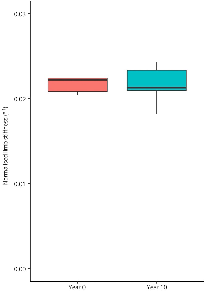
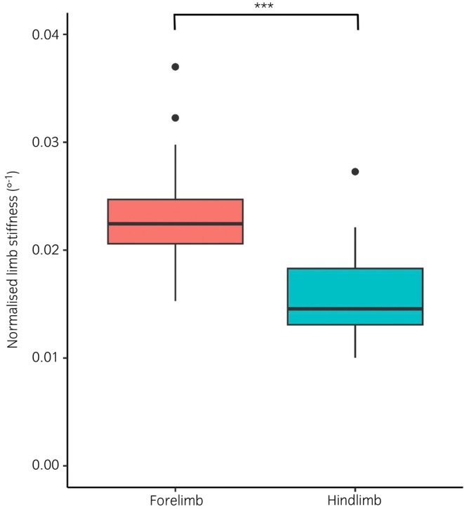
Limb stiffness correlated with weight in forelimbs as shown previously (p < 0.001) but also in hindlimbs (p = 0.006). When normalised to the horse's weight (503 kg, IQR 471.5-560), forelimb stiffness was significantly higher (22.3 [±4.5] × 10 degree) than for the hindlimb (16.4 [±4.0] × 10 degree; p < 0.001). While there were no significant differences between forelimb and hindlimb conformation in unaffected or SDFT injury, both limb stiffness and conformation was significantly greater in limbs with SL injury (p = 0.009 and p = 0.002, respectively).
Small sample size, lack of clinical data including lameness and quantification of injuries.
Injury to the forelimb SDFT does not alter limb stiffness or conformation in the long-term, while hindlimb SL injury simultaneously increases limb stiffness and fetlock angle, suggesting an increase in SL length following injury.
对肢体刚度和形态进行体内测量可提供一种非侵入性的替代评估方法,用于评估指浅屈肌腱(SDFT)和悬韧带(SL)的功能。在此,我们比较了前肢和后肢以及损伤后的情况。
比较前肢和后肢的肢体刚度和形态、随年龄的变化以及SDFT和SL损伤后的情况。
回顾性队列研究。
使用地磅和贴在背侧跗关节上的电子测角仪计算肢体刚度。在肢体负重且抬起对侧肢体(增加负荷)时,同时记录跗关节角度和体重5次。根据角度与负荷回归线的斜率计算双肢的肢体刚度。体重为零时的跗关节角度从图表中推算得出,并用作形态的衡量指标。对未受伤的前肢(n = 42肢)、后肢(n = 19肢)、患有SDFT损伤的前肢(n = 18)和患有SL损伤的后肢(n = 5)进行肢体刚度测量。
如先前所示,前肢的肢体刚度与体重相关(p < 0.001),后肢也如此(p = 0.006)。按马的体重(503 kg,四分位距471.5 - 560)进行标准化后,前肢刚度(22.3 [±4.5]×10度)显著高于后肢(16.4 [±4.0]×10度;p < 0.001)。在未受影响或SDFT损伤的情况下,前肢和后肢形态无显著差异,但在患有SL损伤的肢体中,肢体刚度和形态均显著更大(分别为p = 0.009和p = 0.002)。
样本量小,缺乏包括跛行和损伤量化在内的临床数据。
前肢SDFT损伤长期不会改变肢体刚度或形态,而后肢SL损伤会同时增加肢体刚度和跗关节角度,提示损伤后SL长度增加。