Institute of Diagnostic and Interventional Neuroradiology, Faculty of Medicine and University Hospital Carl Gustav Carus, Technische Universität Dresden, Dresden, Germany.
Department of Pediatrics and Adolescent Medicine, University Medical Center Göttingen, Göttingen, Germany.
Hum Brain Mapp. 2024 Sep;45(13):e70014. doi: 10.1002/hbm.70014.
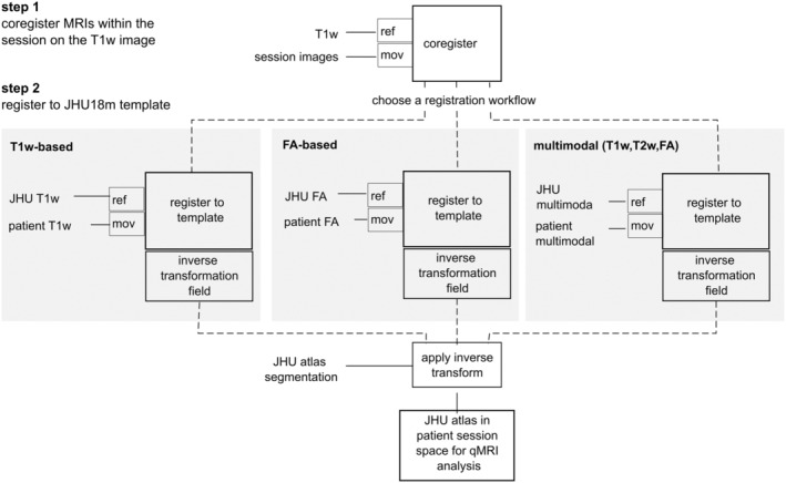
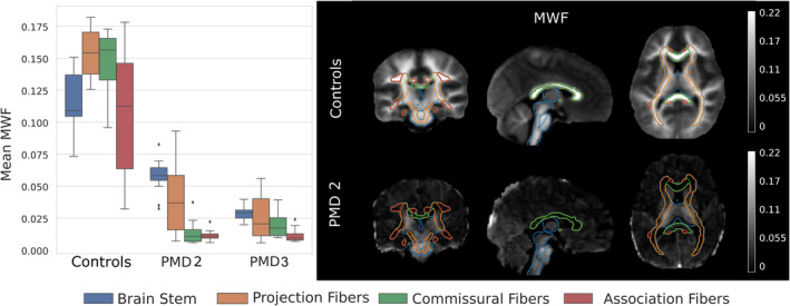
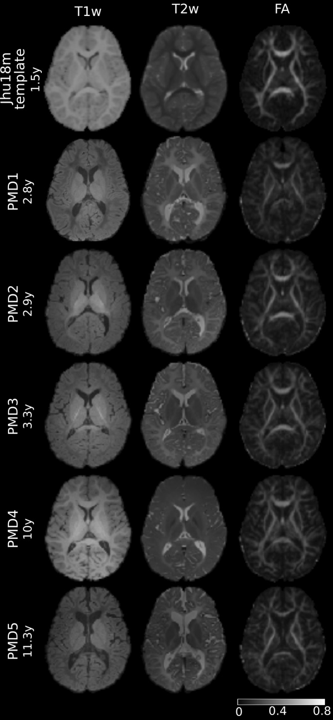
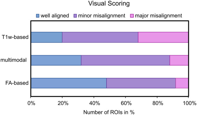
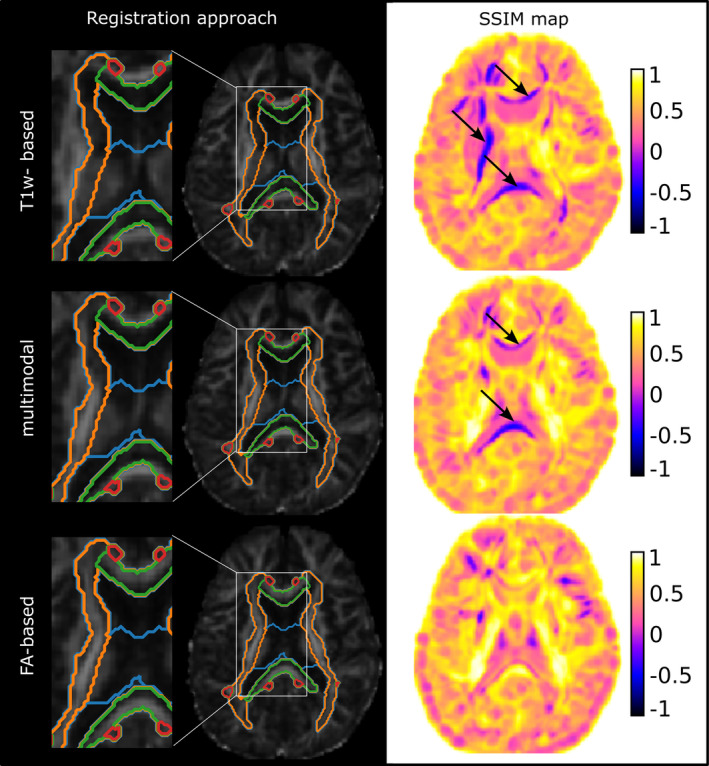
Pelizaeus-Merzbacher disease (PMD) is a rare childhood hypomyelinating leukodystrophy. Quantification of the pronounced myelin deficit and delineation of subtle myelination processes are of high clinical interest. Quantitative magnetic resonance imaging (qMRI) techniques can provide in vivo insights into myelination status, its spatial distribution, and dynamics during brain maturation. They may serve as potential biomarkers to assess the efficacy of myelin-modulating therapies. However, registration techniques for image quantification and statistical comparison of affected pediatric brains, especially those of low or deviant image tissue contrast, with healthy controls are not yet established. This study aimed first to develop and compare postprocessing pipelines for atlas-based quantification of qMRI data in pediatric patients with PMD and evaluate their registration accuracy. Second, to apply an optimized pipeline to investigate spatial myelin deficiency using myelin water imaging (MWI) data from patients with PMD and healthy controls. This retrospective single-center study included five patients with PMD (mean age, 6 years ± 3.8) who underwent conventional brain MRI and diffusion tensor imaging (DTI), with MWI data available for a subset of patients. Three methods of registering PMD images to a pediatric template were investigated. These were based on (a) T1-weighted (T1w) images, (b) fractional anisotropy (FA) maps, and (c) a combination of T1w, T2-weighted, and FA images in a multimodal approach. Registration accuracy was determined by visual inspection and calculated using the structural similarity index method (SSIM). SSIM values for the registration approaches were compared using a t test. Myelin water fraction (MWF) was quantified from MWI data as an assessment of relative myelination. Mean MWF was obtained from two PMDs (mean age, 3.1 years ± 0.3) within four major white matter (WM) pathways of a pediatric atlas and compared to seven healthy controls (mean age, 3 years ± 0.2) using a Mann-Whitney U test. Our results show that visual registration accuracy estimation and computed SSIM were highest for FA-based registration, followed by multimodal, and T1w-based registration (SSIM = 0.67 ± 0.04 vs. SSIM = 0.60 ± 0.03 vs. SSIM = 0.40 ± 0.14). Mean MWF of patients with PMD within the WM pathways was significantly lower than in healthy controls MWF = 0.0267 ± 0.021 vs. MWF = 0.1299 ± 0.039. Specifically, MWF was measurable in brain structures known to be myelinated at birth (brainstem) or postnatally (projection fibers) but was scarcely detectable in other brain regions (commissural and association fibers). Taken together, our results indicate that registration accuracy was highest with an FA-based registration pipeline, providing an alternative to conventional T1w-based registration approaches in the case of hypomyelinating leukodystrophies missing normative intrinsic tissue contrasts. The applied atlas-based analysis of MWF data revealed that the extent of spatial myelin deficiency in patients with PMD was most pronounced in commissural and association and to a lesser degree in brainstem and projection pathways.
佩利兹-梅茨巴赫病(PMD)是一种罕见的儿童脑白质营养不良。定量评估明显的髓鞘缺失并描绘细微的髓鞘形成过程具有重要的临床意义。定量磁共振成像(qMRI)技术可以提供关于髓鞘状态、其空间分布和脑成熟过程中动态变化的体内见解。它们可能成为评估髓鞘调节治疗效果的潜在生物标志物。然而,针对受影响儿科脑的图像量化和统计比较的配准技术,特别是那些图像组织对比度低或异常的脑,与健康对照组相比,尚未建立。本研究的目的首先是开发和比较基于图谱的 PMD 患儿 qMRI 数据定量分析的后处理管道,并评估其配准准确性。其次,应用优化的管道,使用 PMD 患者和健康对照组的髓鞘水成像(MWI)数据来研究空间性髓鞘缺失。这是一项回顾性单中心研究,纳入了 5 名 PMD 患者(平均年龄 6 岁±3.8 岁),他们接受了常规脑 MRI 和弥散张量成像(DTI)检查,部分患者还进行了 MWI 检查。研究了三种将 PMD 图像与儿科模板配准的方法。这些方法基于(a)T1 加权(T1w)图像,(b)各向异性分数(FA)图,和(c)T1w、T2 加权和 FA 多模态组合。通过视觉检查和结构相似性指数法(SSIM)计算来确定配准准确性。使用 t 检验比较了不同配准方法的 SSIM 值。从 MWI 数据中量化髓鞘水分数(MWF),作为相对髓鞘化的评估指标。从四个儿科图谱的四个主要白质(WM)通路中的两个 PMD(平均年龄 3.1 岁±0.3 岁)中获得平均 MWF,并与七个健康对照组(平均年龄 3 岁±0.2 岁)进行比较,使用 Mann-Whitney U 检验。我们的结果表明,FA 配准的视觉配准准确性估计和计算的 SSIM 值最高,其次是多模态配准和 T1w 配准(SSIM=0.67±0.04 比 SSIM=0.60±0.03 比 SSIM=0.40±0.14)。PMD 患者在 WM 通路中的平均 MWF 明显低于健康对照组的 MWF=0.0267±0.021 比 MWF=0.1299±0.039。具体来说,MWF 可在已知在出生时(脑干)或出生后(投射纤维)髓鞘化的脑结构中测量,但在其他脑区(连合纤维和联络纤维)中几乎不可测量。总之,我们的结果表明,FA 配准管道的配准准确性最高,为缺乏正常固有组织对比的脑白质营养不良提供了替代传统 T1w 配准方法的选择。应用基于图谱的 MWF 数据分析显示,PMD 患者的空间性髓鞘缺失程度在连合纤维和联络纤维中最为明显,在脑干和投射纤维中则相对较轻。