Alayyan Amin, Hammad Tarek, Majeed Salman
Internal Medicine, Northampton General Hospital NHS Trust, Northampton, GBR.
Cardiology, Northampton General Hospital NHS Trust, Northampton, GBR.
Cureus. 2024 Sep 4;16(9):e68654. doi: 10.7759/cureus.68654. eCollection 2024 Sep.
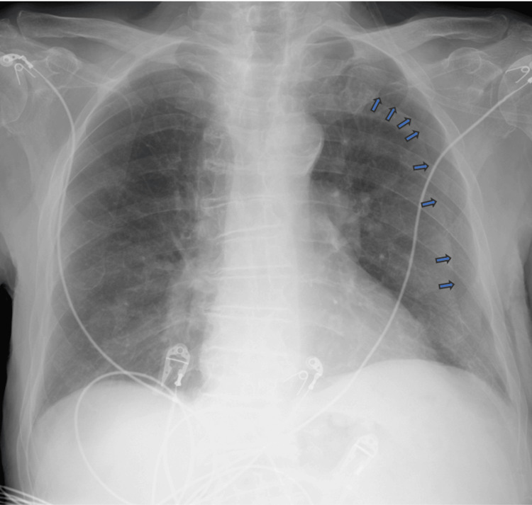
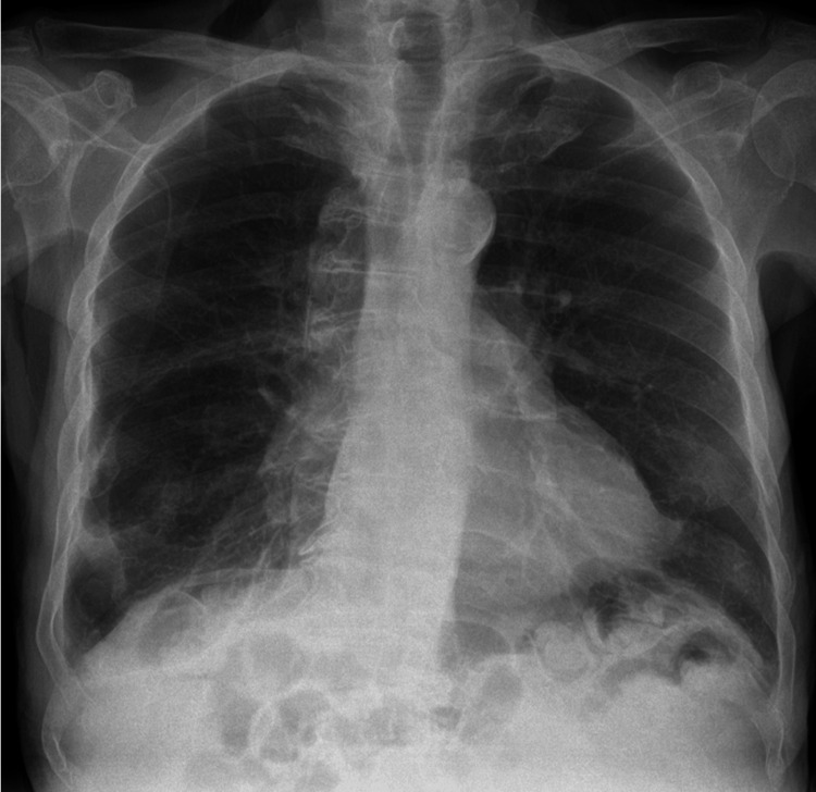
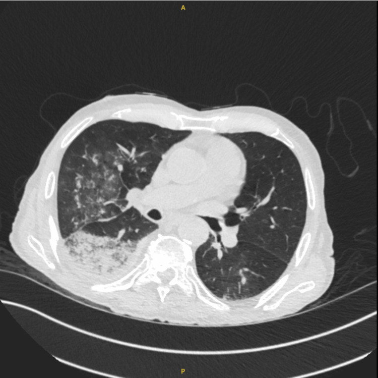
We present the case of a male patient in his late 80s who presented with a fall with symptoms and signs of community-acquired pneumonia. Chest X-ray showed the suspicion of a left-sided pneumothorax. A CT of the chest subsequently ruled out the presence of a pneumothorax on the left side. The pseudo-pneumothorax on the chest X-ray was secondary to a skinfold. This case highlights how well a skinfold can mimic pneumothorax. Careful clinical and radiological examination with bedside lung ultrasound and/or CT of the chest can help differentiate true pneumothorax from pseudo-pneumothorax, provided the patient is hemodynamically stable. Our case highlights the importance of clinical examination, various imaging modalities, and confirmation of a diagnosis before proceeding to interventional procedures in the context of limited clinical suspicion of the differential.
我们报告了一例80多岁男性患者的病例,该患者因跌倒就诊,伴有社区获得性肺炎的症状和体征。胸部X线显示怀疑左侧气胸。随后的胸部CT排除了左侧气胸的存在。胸部X线片上的假性气胸继发于皮肤褶皱。该病例突出了皮肤褶皱模拟气胸的程度。如果患者血流动力学稳定,仔细的临床和放射学检查,结合床边肺部超声和/或胸部CT,有助于区分真性气胸和假性气胸。我们的病例强调了在对鉴别诊断临床怀疑有限的情况下,临床检查、各种成像方式以及在进行介入程序之前确诊的重要性。