Department of Systems Biology, School of Life Sciences, Guangdong Provincial Key Laboratory of Cell Microenvironment and Disease Research, Shenzhen Key Laboratory of Cell Microenvironment, Southern University of Science and Technology, Shenzhen, 518055, China.
Department of Pathology, School of Medicine and University of Pittsburgh Cancer Institute, University of Pittsburgh, Pittsburgh, PA, 15260, USA.
Cell Mol Life Sci. 2024 Sep 5;81(1):385. doi: 10.1007/s00018-024-05422-w.
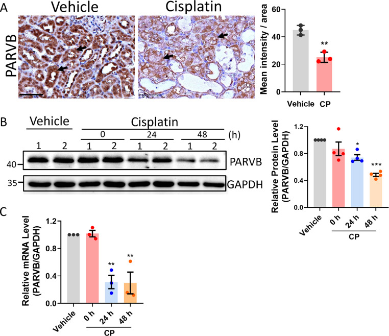
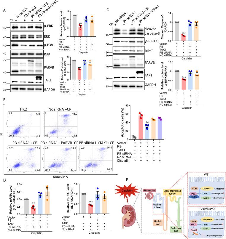
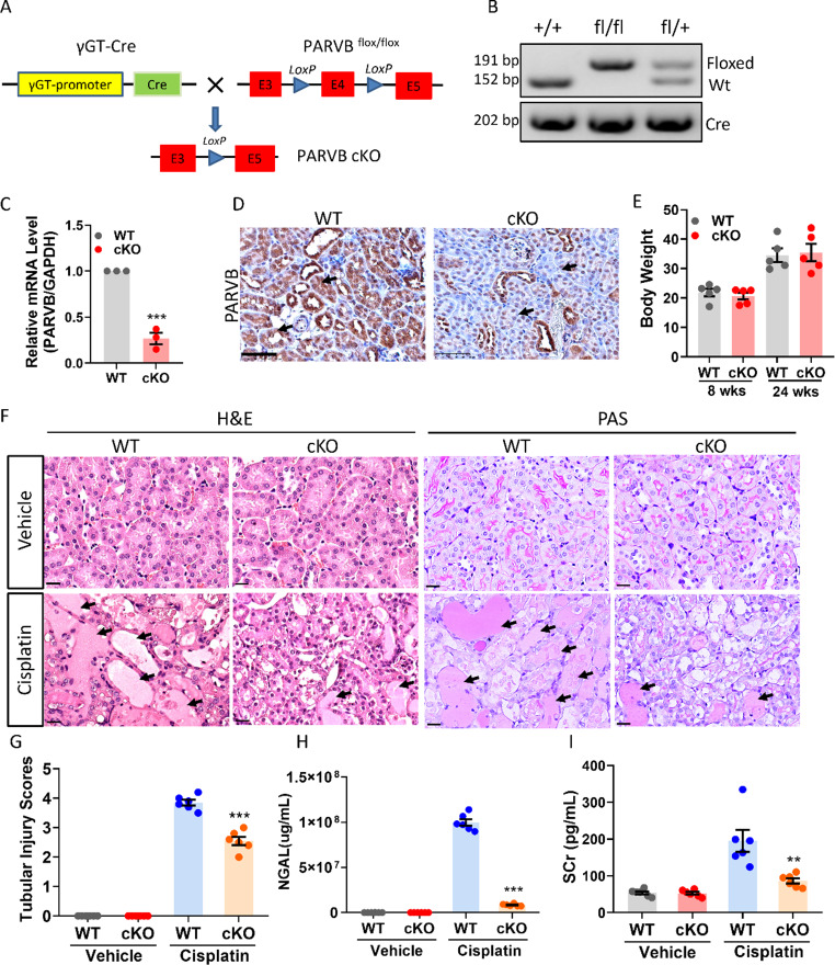
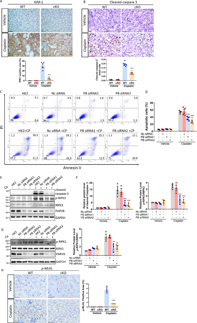
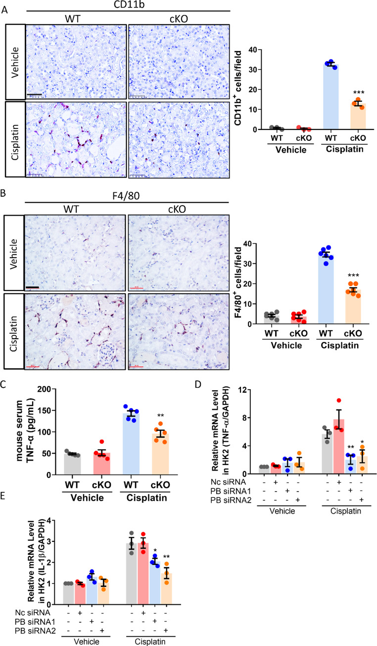
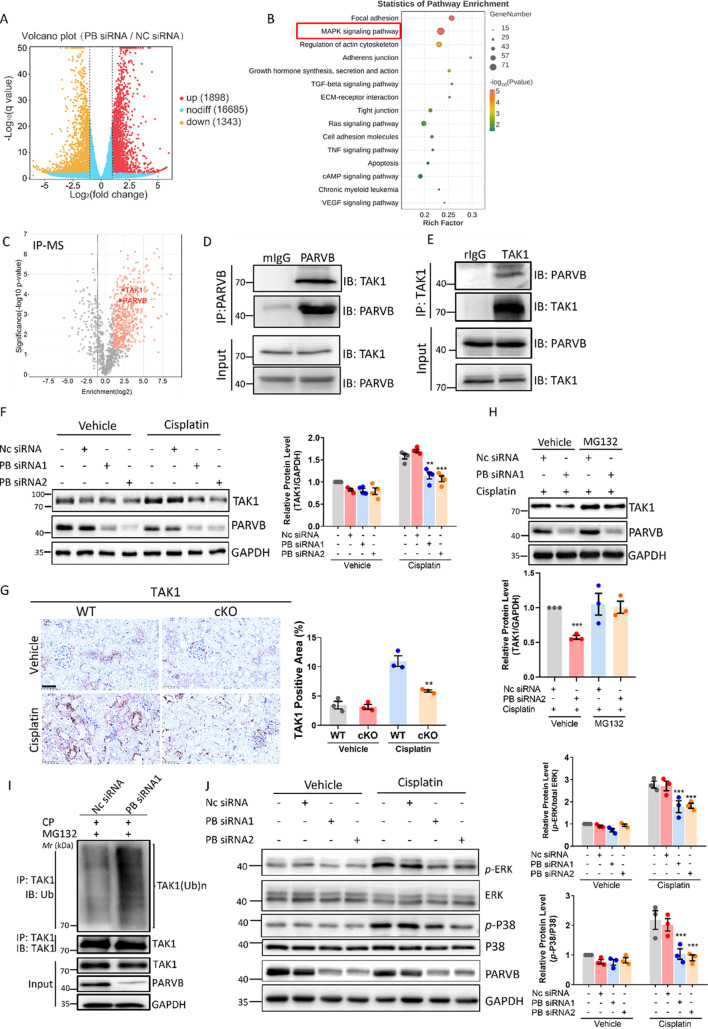
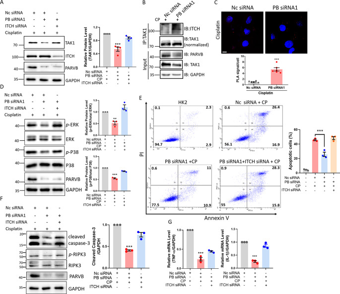
Cisplatin-induced renal tubular injury largely restricts the wide-spread usage of cisplatin in the treatment of malignancies. Identifying the key signaling pathways that regulate cisplatin-induced renal tubular injury is thus clinically important. PARVB, a focal adhesion protein, plays a crucial role in tumorigenesis. However, the function of PARVB in kidney disease is largely unknown. To investigate whether and how PARVB contributes to cisplatin-induced renal tubular injury, a mouse model (PARVB cKO) was generated in which PARVB gene was specifically deleted from proximal tubular epithelial cells using the Cre-LoxP system. In this study, we found depletion of PARVB in proximal tubular epithelial cells significantly attenuates cisplatin-induced renal tubular injury, including tubular cell death and inflammation. Mechanistically, PARVB associates with transforming growth factor-β-activated kinase 1 (TAK1), a central regulator of cell survival and inflammation that is critically involved in mediating cisplatin-induced renal tubular injury. Depletion of PARVB promotes cisplatin-induced TAK1 degradation, inhibits TAK1 downstream signaling, and ultimately alleviates cisplatin-induced tubular cell damage. Restoration of PARVB or TAK1 in PARVB-deficient cells aggravates cisplatin-induced tubular cell injury. Finally, we demonstrated that PARVB regulates TAK1 protein expression through an E3 ligase ITCH-dependent pathway. PARVB prevents ITCH association with TAK1 to block its ubiquitination. Our study reveals that PARVB deficiency protects against cisplatin-induced tubular injury through regulation of TAK1 signaling and indicates targeting this pathway may provide a novel therapeutic strategy to alleviate cisplatin-induced kidney damage.
顺铂诱导的肾小管损伤在很大程度上限制了顺铂在恶性肿瘤治疗中的广泛应用。因此,鉴定调控顺铂诱导的肾小管损伤的关键信号通路具有重要的临床意义。PARVB 是一种黏着斑蛋白,在肿瘤发生中起着关键作用。然而,PARVB 在肾脏疾病中的功能在很大程度上是未知的。为了研究 PARVB 是否以及如何参与顺铂诱导的肾小管损伤,我们使用 Cre-LoxP 系统在近端肾小管上皮细胞中特异性敲除 PARVB 基因,构建了 PARVB 敲除小鼠模型(PARVB cKO)。在这项研究中,我们发现近端肾小管上皮细胞中 PARVB 的缺失显著减轻了顺铂诱导的肾小管损伤,包括肾小管细胞死亡和炎症。机制上,PARVB 与转化生长因子-β激活激酶 1(TAK1)相关,TAK1 是细胞存活和炎症的中央调节因子,在介导顺铂诱导的肾小管损伤中起着关键作用。PARVB 的缺失促进了顺铂诱导的 TAK1 降解,抑制了 TAK1 下游信号转导,最终减轻了顺铂诱导的肾小管细胞损伤。在 PARVB 缺陷细胞中恢复 PARVB 或 TAK1 会加重顺铂诱导的肾小管细胞损伤。最后,我们证明 PARVB 通过 E3 连接酶 ITCH 依赖性途径调节 TAK1 蛋白表达。PARVB 阻止 ITCH 与 TAK1 结合,从而阻止其泛素化。我们的研究揭示了 PARVB 通过调节 TAK1 信号来保护顺铂诱导的肾小管损伤,并表明靶向该途径可能为减轻顺铂诱导的肾脏损伤提供一种新的治疗策略。