Deshmukh Asawari, Deshmukh Sanika
Radiology and Fetal Medicine, Dhruv Diagnostic and Imaging Clinic, Nagpur, IND.
Radiodiagnosis, Dr. D. Y. Patil Medical College, Hospital and Research Centre, Pune, IND.
Cureus. 2024 Aug 8;16(8):e66462. doi: 10.7759/cureus.66462. eCollection 2024 Aug.
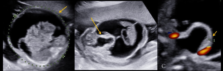
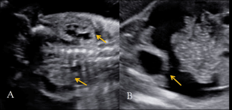
Prenatal ultrasonography (USG) plays a crucial role in diagnosing fetal urinary tract anomalies and distinguishing between lower urinary tract obstructive (LUTO) and neurological causes (seen with spinal dysraphism, myelomeningocele, meningocele, and sacral agenesis) of urinary bladder distension. Fetal urinary ascites, a rare but severe complication, can result from bladder rupture associated with obstructive uropathy such as posterior urethral valves (PUV). This case study presents a rare instance of fetal urinary ascites due to PUV detected during prenatal ultrasonography at 20 weeks of gestation (WOG). By highlighting this uncommon but clinically significant condition, we aim to enhance the understanding and management of similar cases in clinical practice.
产前超声检查(USG)在诊断胎儿泌尿系统异常以及区分下尿路梗阻(LUTO)和膀胱扩张的神经学原因(如脊柱裂、脊髓脊膜膨出、脊膜膨出和骶骨发育不全)方面起着关键作用。胎儿尿腹水是一种罕见但严重的并发症,可由与诸如后尿道瓣膜(PUV)等梗阻性尿路病相关的膀胱破裂引起。本病例研究呈现了一例罕见的因妊娠20周(WOG)产前超声检查时发现的后尿道瓣膜导致的胎儿尿腹水病例。通过强调这种罕见但具有临床意义的情况,我们旨在提高临床实践中对类似病例的认识和管理水平。